Original Article Tumor-Associated Macrophages Derived TGF-β‒Induced Epithelial to Mesenchymal Transition in Colorectal Cancer Cells through Smad2,3-4/Snail Signaling Pathway
Cancer Research and Treatment : Official Journal of Korean Cancer Association 2019;51(1):252-266.
DOI: https://doi.org/10.4143/crt.2017.613
Published online: April 25, 2018
Department of Colorectal and Anal Surgery, the First Affiliated Hospital of Wenzhou Medical University, Wenzhou, China
Correspondence: Xiangbin Wu, MD Department of Colorectal and Anal Surgery, the First Affiliated Hospital of Wenzhou Medical University, Nanbaixiang, Ouhai District, Wenzhou, Zhejiang 325000, China Tel: 86-0577-13806897120 Fax: 86-0577-13806897120 E-mail: wxb113@126.com
• Received: December 29, 2017 • Accepted: April 24, 2018
This is an Open Access article distributed under the terms of the Creative Commons Attribution Non-Commercial License (http://creativecommons.org/licenses/by-nc/4.0/) which permits unrestricted non-commercial use, distribution, and reproduction in any medium, provided the original work is properly cited.
We investigated the role of tumor-associated macrophages (TAMs) on the epithelial to mesenchymal transition (EMT) of colorectal cancer cells and determined the potential mechanism involved in the metastatic process.
Materials and Methods
In this study, flow cytometry was used to detect the expression of target proteins. We used transwell assay to evaluate the migration of cancer cells under specific conditions. Using real-time polymerase chain reaction, we examined the expressions of cytokines and EMT-related markers in mRNA level. Animal assay was performed for analysis in vivo and hematoxylin and eosin was used to visualize the effect of TAMs on tumor metastasis. We also used immunohistochemistry and Western blotting to detect the expression of target proteins.
Results
Here, we observed enrichment of TAMs in colorectal tumor tissues, resulting in high metastasis in clinical therapy. Moreover, those TAMs could facilitate the EMT progression of colorectal cancer cells, which is induced by the transforming growth factor-β (TGF-β) derived from TAMs, leading to the invasion and migration of cancer cells.
Conclusion
Our results demonstrated that TAMs contributed the EMT progression through a TGF-β/Smad2,3-4/Snail signaling pathway, and disrupting this pathway with TGF-β receptor inhibitor could suppress metastasis, readjusting our focus to the connection of TAMs and cancer metastasis.
Colorectal carcinoma is the second most common cause of cancer-associated death worldwide [1,2]. Though half of colorectal cancer patients showed a positive initial response that benefited from the successful surgery or multimodal treatment [3], many patients had worse treatment efficiency and dismal prognosis which were considered to be induced by visceral metastasis [3]. Moreover, the 5-year survival for patients with metastasis was lower than those who had no metastasis [4]. However, the specific molecular mechanisms underlying the development of metastasis in colorectal cancer remain unclear [5]. Hence, identifying the drivers that influence the distant metastasis may lay an important foundation to make progress in the diagnosis and therapy for colorectal cancer patients.
Given the potential role on clinical treatment, a better understanding of the mechanism of distant metastases in colorectal cancer is critical. Increasing evidences reveals cancer metastasis depends on various curial factors, including up-regulation of integrins [6] and E-cadherin [7,8], epithelial–mesenchymal transition (EMT) progression of tumor cells [9], hypoxia [10], tumor microenvironment [11] and so on [12]. EMT was initially reported as a tightly regulated lineage change during gastrulation, neural crest delamination, heart valve formation, and embryonic development. Recently, increasing attention has been attracted for the important role of EMT in cancer metastasis [13]. A lot of molecules are responsible for the EMT in the tumor cells, including inflammatory cytokines, growth factors and numerous transcription factors. In recent years, the tumor microenvironment has received attention as an important determination of cancer cells metastasis, among which tumor-associated macrophages (TAMs) have been reported to play a prominent functional role in cancer behaviors [14]. TAMs are the major inflammatory component in many tumor microenvironments, which affect different aspects of tumor behaviors [15], including participation in tumor growth in several ways, such as transforming growth factor-β (TGF-β) secretion [16], remodeling of tumor microenvironment, inflammation suppression [17] and so on. TGF-β is one of the major endogenous regulators of cell growth, which shows enhanced expression in various human cancers, including breast cancer, colorectal cancer, pancreatic cancer and so on [18]. TGF-β participates in multifarious tumor progress, including potent immunosuppressive effects, EMT progression of tumor cells [19]. Increasing evidences demonstrated that TAMs facilitate distant metastasis in various tumors [20], including lung cancers, breast cancer and so on. However, the specific mechanism of metastasis induced by TAMs is still unclear and it is a challenge to inhibit the metastasis of colorectal cancer in clinic therapy.
In our study, our aim is to reveal the expressional profile of TAMs in the colorectal cancers and investigate its potential role in the metastatic progress. Moreover, finding the underlying mechanism involved in the colorectal cancer metastasis may lay an important foundation for the clinical treatment. We examined enriched TAMs in tumor tissues from colorectal patients with high metastasis, which was proved to facilitate tumor cells migration and neoplasm metastasis. Furthermore, our study reveals that TAMs promote the progression of EMT in colorectal cancer cells through the secretion of TGF-β, resulting in the lung metastasis. TGF-β regulates the EMT progression through activation of Smad2,3-4/Snail/E-cadherin signaling pathway. Blockade of TGF-β/TGF-β receptor synapse with TGF-β receptor inhibitor suppresses colorectal cancer cells lung metastasis.
Materials and Methods
1. Patient samples
Colorectal cancer samples were sterilely obtained after the surgery at the first affiliated hospital of Wenzhou Medical University and were sent to the laboratory within 2 hours. According to the clinical data, samples were divided into metastasis and non-metastasis groups. The clinical information was listed in Table 1. All samples were confirmed as colorectal cancer by a pathologist expert.
2. Cell lines and reagents
HCT116 (human colorectal cancer cell line) and CT26 (mouse colorectal cancer cell line) cells were purchased from the Cell Bank of Chinese Academy of Sciences (Shanghai, China). All cell lines were cultured in RPMI-1640 (Invitrogen, Carlsbad, CA) supplemented with 10% fetal bovine serum (Gibco, Grand Island, NY), penicillin (100 U/mL), and streptomycin (0.1 mg/mL). Recombinant interleukin (IL)-10, TGF-β1, CCL21, CCL18, CXCL12, and vascular endothelial growth factor (VEGF) proteins were purchased from Yanjing Company (Shanghai, China). TGF-β1 neutralization antibody was purchased from Abcam (Cambridge, UK). Clodronate liposomes were purchased from Yisheng Company (Yu Yao, China). LY2109761 was purchased from Selleck (Houston, TX). Anti-CCL2 and anti-mouse CSF1R (CD115) were purchased from Bio X Cell (Lebanon, NH).
3. TAMs preparation and collection from tumor tissues
For mouse TAMs, BALB/c mice (females, 6-8 weeks old) were purchased from Vital River Laboratory Animal Technology Company (Beijing, China). Mice were implanted subcutaneously with 1×10⁵ CT26 cells into the right flank. Mice were scarified and separated the tumor tissues when the tumor reached to 7 mm×7 mm. After digesting the tissues by using collagenase II and IV in 37°C for 2 hours, TAMs were obtained by using separating medium (Junrui Techonology, Shenzhen, China) and sorted with Anti-CD45 and Anti-F4/80 Microbeads (Miltenyi Biotec, Cologne, Germany). For human TAMs, single cell from tumor samples were harvested with DNase and collagenase II and IV digestion in 37°C for 2 hours. TAMs were collected by CD45+CD68+ sorting (eBioscience, San Diego, CA).
4. Lung metastasis assay
BALB/c mice were implanted subcutaneously with 1×10⁵ CT26 cells into the right flank. Twenty-five days later, mice were sacrificed and the lungs were harvested to measure lung weight and examine the number of pulmonary nodules [7]. To further measure the metastasis in lung, the sections were also stained with hematoxylin and eosin (H&E) (Solarbio, Beijing, China).
5. Immunohistochemistry
After sacrificing mice, tissues were kept in 4% paraformaldehyde (PFA) overnight, then the samples were processed, embedded in paraffin, and sectioned at 4 μm for further study. To detect the distribution of CD68 or TGF-β in human colorectal cancer tissues, 10 metastatic samples and 10 non-metastatic samples were conducted by immunohistochemistry (IHC). Antigen retrieval was done using citric acid and sodium citrate in a Microwave oven (Media, Guangdong, China). Then the sections were incubated with CD68 (1:500, Abcam) or TGF-β (1:500, Abcam) at 4°C overnight, followed by signal amplification using a ABC HRP Kit (Thermo Fisher, Waltham, MA) and counter-staining with hematoxylin, dehydration with series of graded ethanol and cleaned with xylene [1]. Microscope (Leica, Barnack, Oskar, Germany) was used to visualize the sections. Ten visions were taken from each sample randomly for intensity analysis and one representative graph were presented.
6. Immunofluorescence
Samples were blocked in 5% bovine serum albumin in phosphate buffered saline (PBS) for 1 hour, E-cadherin (1:200, Abcam) and vimentin (1:300, Abcam) were incubated at 4°C overnight, followed by signal amplification using TSA Kit (PerkinElmer, Waltham, MA) [2]. An Olympus confocal microscope (Tokyo, Japan) was used to visualize.
7. Western blotting
Samples were solubilized with an equal volume of loading buffer (125 mM Tris-HCl, pH 6.8, 4% sodium dodecyl sulfate, 20% glycerol, 0.05% bromophenolblue, 5% β-mercaptoethanol) and were boiled for 10 minutes, then samples were separated by sodium dodecyl sulfate polyacrylamide gel electrophoresis, followed by transferring to polyvinylidene difluoride membranes and detecting by immunoblotting with primary antibodies against E-cadherin (1:500, Abcam), vimentin (1:500, Abcam), p-Smad2 (1:500, Abcam), p-Smad3 (1:500, Abcam), Smad4 (1:500, Abcam), and Snail (1:500, Abcam), respectively at 4°C overnight. Then HRP-conjugated secondary antibody (CST, Boston, MA) was incubated for 1 hour at room temperature, and visualized by using ECL detection kit (CST) [3]. β-Actin (1:1,000, Abcam) was used as an internal control.
8. Real-time polymerase chain reaction
The quantification of gene transcripts was performed by real-time polymerase chain reaction (PCR) using SYBR green dye (Solarbio) [4]. Glyceraldehyde 3-phosphate dehydrogenase (GAPDH) was used for normalization. The primers used are listed as follows: human GAPDH forward primer 5'-GGAGCGAGATCCCTCCAAAAT-3', reverse primer 5'-GGCTGTTGTCATACTTCTCATGG-3'; human E-cadherin, forward primer 5'-CGAGAGCTACACGTTCACGG-3', reverse primer 5'-GGGTGTCGAGGGAAAAATAGG-3'; human vimentin forward primer 5'-GACG CCATCAACACCGAGTT-3', reverse primer 5'-CTTTGTCGTTGGTTAGCTGGT-3'; human Tgf b1 forward primer 5'- GGCCAGATCCTGTCCAAGC-3', reverse primer 5'-GTGGGTTTCCACCATTAGCAC-3'; human Tgf b receptor forward primer 5'-GTAGCTCTGATGAGTGCAATGAC-3', reverse primer 5'-CAGATATGGCAACTCCAGTG-3'; human IL-10 forward primer 5'-GACTTTAAGGGTTACCTGGGTTG-3', reverse primer 5'-TCACATGCGCCTTGATGTCTG-3'; human IL-10 receptor forward primer 5'-CCTCCGTCTGTGTGGTTTGAA-3', reverse primer 5'-CACTG CGGTAAGGTCATAGGA-3'; human VEGFα forward primer 5'-AGGGCAGAATCATCACGAAGT-3', reverse primer 5'-AGGGTCTCGATTGGATGGCA-3'; human VEFGR forward primer 5'-AGGGCAGAATCATCACGAAGT-3', reverse primer 5'-AGGGTCTCGATTGGATGGCA-3'; human CCL17 forward primer 5'- CGGGACTACCTGGGACCTC-3', reverse primer 5'-CCTCACTGTGGCTCTTCTTCG-3'; human CCR4 forward primer 5'-CCCACGGATATAGCAGACACC-3', reverse primer 5'-GTGCAAGGCTTGGGGATACT -3'; human CCL21 forward primer 5'-GTTGCCTCAAGTACAGCCAAA-3', reverse primer 5'-AGAACAGGATAGCTGGGATGG-3'; human CCR7 forward primer 5'-TGAGGTCACGGACGATTACAT-3', reverse primer 5'-GTAGGCCCACGAAACAAATGAT-3'; human CXCL12 forward primer 5'-ATTCTCAACACTCCAAACTGTGC-3', reverse primer 5'-ACTTTAGCTTCGGGTCAATGC-3'; human CXCR4 forward primer 5'-ACTACACCGAGGAAATGGGCT-3', reverse primer 5'-CCCACAATGCCAGTTAAGAAGA-3'; mouse GAPDH forward primer 5'-AGGTCGGTGTGAACGGATTTG-3', reverse primer 5'-GGGGTCGTTGATGGCAACA-3'; mouse E-cadherin, forward primer 5'-CAGTTCCGAGGTCTACACCTT-3', reverse primer 5'-TGAATCGGGAGTCTTCCGAAAA-3'; mouse vimentin forward primer 5'-CGTCCACACGCACCTACAG-3', reverse primer 5'-GGGGGATGAGGAATAGAGGCT-3'; mouse Tgfb1 forward primer 5'-CCACCTGCAAGACCATCGAC-3', reverse primer 5'-CTGGCGAGCCTTAGTTTGGAC-3'; mouse Tgfb receptor forward primer 5'-AGTCGGATGTGGAAATGGAAG-3', reverse primer 5'-CTGGCCATGACATCACTGTTA-3'; mouse IL-10 forward primer 5'-CTTACTGACTGGCATGAGGATCA-3', reverse primer 5'-GCAGCTCTAGGAGCATGTGG-3'; mouse IL-10 receptor forward primer 5'-TTGTCGCGTTTGCTCCCATT-3', reverse primer 5'-GAAGGGCTTGGCAGTTCTG-3'; mouse VEGFα forward primer 5'-CTGCCGTCCGATTGAGACC-3', reverse primer 5'-CCCCTCCTTGTACCACTGTC-3'; mouse VEGFα receptor forward primer 5'-TTTGGCAAATACAACCCTTCAGA-3', reverse primer 5'-GCTC CAGTATCATTTCCAACCA-3'; mouse CCL17 forward primer 5'-TACCATGAGGTCACTTCAGATGC-3', reverse primer 5'-GCACTCTCGGCCTACATTGG-3'; mouse CCR4 forward primer 5'-TGCACCAAGGAAGGTATCAAGG-3', reverse primer 5'-GTACACGTCCGTCATGGACTT-3'; mouse CCL21 forward primer 5'-GTGATGGAGGGGGTCAGGA-3', reverse primer 5'-GGGATGGGACAGCCTAAACT-3'; mouse CCR7 forward primer 5'-CAGGTGTGCTTCTGCCAAGAT-3', reverse primer 5'-GGTAGGTATCCGTCATGGTCT-3'; mouse CXCL12 forward primer 5'-TGCATCAGTGACGGTAAACCA-3', reverse primer 5'-CACAGTTTGGAGTGTTGAGGAT-3'; mouse CXCR4 forward primer 5'-GACTGGCATAGTCGGCAATG-3', reverse primer 5'-AGAAGGGGAGTGTGATGACAAA-3'.
9. Cell invasion assay
For invasion assay, 30 μg matrigel (BD Biosciences, FranklinLakes, NJ) was used to coat the upper chamber of 24 wells plates (8 μm, Corning Company, Corning, NY). Then, CT26 or HCT116 cells (3×10⁴) suspended in fetal calf serum (FCS)‒free 1640 RPMI medium were seeded in the upper chamber, meanwhile, the lower chamber was added with macrophage culturing supernatants. RPMI-1640 without FCS was used as the negative control. Cells were allowed to migrate at 37°C for 18 hours. Then 4% PFA was used to fix the cells that invaded the lower surface of the membrane, followed by 1% crystal violet staining for calculation [5]. In some experiments, the lower chamber was added with the following recombinant proteins, IL-10/TGF-β/CCL21/CCL18/CXCL12/VEGF, to determining whether they involve in the cell invasion process.
10. Wound healing assay
For wound healing assay, CT26 or HCT116 cells were seeded in 6-well plates, a 10-μL microtiter tip was used to cut trace longitudinally when cells reaching 85% confluent, then cells were treated with or without macrophage culturing supernatants or TGF-β for 24 hours [6]. Using the microscope, pictures were taken as 200× for analysis.
11. RNA interfering
For Snail knockdown in tumor cells, 2×10⁵ CT26 cells/well were seeded in a six-well plate and starved in an antibioticfree growth medium for 24 hours before transfection. Snail siRNA (4 mL, 0.5 mg) or a mock siRNA solution was performed for 48 hours according to the manufacturer’s protocol. The transfection efficacy was validated by western blot [9]. The primers were designed and constructed by Gene Pharma Company (South San Francisco, CA). Then the cells were treated with TGF-β to examine the cell morphology in microscope. The following sequences were successfully: siRNA-Snail sense 5'-GAT CCG CCT AAC TAC AGC GAG CTG TTC AAG AGA CAG CTC GCT GTA GTT AGG CTT TTT TGG AAA-3' and antisense 5'-AGC TTT TCC AAA AAA GCC TAA CTA CAG CGA GCT GTC TCT TGA ACA GCT CGC TGT AGT TAG GCG-3'; the mock vector sense 5'-GAT CCG TAT TGC CTA GCA TTA CGT TTC AAG AGA ACG TAA TGC TAG GCA ATA CTT TTT TGG AAA-3' and antisense 5'-AGC TTT TCC AAA GTA TTG CCT AGC ATT ACG TTC TCT TGA AAC GTA ATG CTA GGC AAT ACG-3'.
12. Flow cytometry
To examine the percentage of macrophage in patients, cells were incubated with CD68 (eBioscience) for 20 minutes at room temperature, after washed twice and then re-suspended in PBS [10]. For intracellular TGF-β (eBioscience) analysis, mice TAMs and peritoneal macrophages or human TAMs and peripheral blood monocytes were collected and fixed, after permeabilization, anti-human or anti-mouse TGF-β were stained respectively. Flow cytometry was performed on the BD Canto II (BD Biosciences). 7-AAD was used to exclude the dead cells. IgG (Abcam) was used as the negative control.
13. Animal protocols and treatments
To evaluate the effects of TGF-β on metastasis in colorectal cancer, 5×10⁵ CT26 were subcutaneous injected into the BALB/c mice (female, 6-8 weeks, Huafukang, Shenzheng, China). On day 15, mice were treated with PBS, TGF-β (250 ng), TAMs cultural supernatant and TAMs cultural supernatant combined with TGF-β neutralizing antibody (200 μg) by intratumoral injection. Mice were sacrificed for the lung metastasis analysis on day 25. To evaluate the anti-metastasis effects of LY2109761, 5×10⁵ CT26 were subcutaneous injected into the BALB/c mice. On day 15, 18, and 21, mice were treated with PBS or LY2109761 (50 mg/kg) by intratumoral injection. On day 25, the mice were sacrificed for the lung metastasis analysis.
14. Enzyme-linked immunosorbent assay
The concentrations of TGF-β in tumor tissues from metastatic and non-metastatic patients were quantified by using commercially available enzyme-linked immunosorbent assay (ELISA) Kits (Dakewe Biotech Co., Ltd., Shenzhen, China). Assays were performed according to the manufacturer’s protocol and read at 450 nm by using a microplate reader (Thermo Scientific).
15. Statistical analysis
All data were represented as mean±SEM. GraphPad Prism ver. 6.0 software was used to analyze data. Specific statistical tests used were paired and unpaired t tests. p-value < 0.05 were considered statistically significant.
16. Ethical statement
All subjects gave written informed consent. Ethical approval was obtained from the Committee of the First Affiliated Hospital of Wenzhou Medical University.
Results
1. TAMs facilitate colorectal cancer cells metastasis
Recently, TAMs accumulation level has been shown to be of significant in cancer cells metastasis in a variety of cancers. To test the infiltration level of TAMs in colorectal cancer, we analyzed CD68+ cells in CD45+ cells in metastatic (patients/M) and non-metastatic (patients/N) colorectal cancer samples from clinical patients by flow cytometry. Interestingly, we found that metastatic patients have more TAMs accumulated than non-metastatic patients in tumor tissues (Fig. 1A). Moreover, we verified this by IHC and the CD68 IHC intensity was correlated with flow cytometry analysis (Fig. 1B). It has been reported that the accumulated TAMs in breast cancer and lung cancer could induce the metastasis. Our initial results suggested that colorectal cancer tissues have TAMs accumulation and the infiltration level was much higher in metastatic patients’ tissues than non-metastatic patients’ (Fig. 1A and B). To clarify whether TAMs could promote the colorectal cancer metastasis, the CT26 mouse subcutaneous model was established. We injected 105 macrophages which was isolated from tumor tissues into tumor site. On the day 25, mice were sacrificed, lungs were weighed, and the metastatic pulmonary tumor nodules on the surface of lungs were counted. We found that the macrophages injection increased the lung weight compared with PBS injection group (Fig. 1C) and increased the pulmonary tumor nodules in lung as well (Fig. 1D). Further, we observed that CT26 cells seeded more lung metastasis with macrophages injection, which was confirmed by the whole lung H&E staining (Fig. 1E). These results indicate that TAMs could promote the colorectal cancer cells lung metastatic seeding.
To further determine whether TAMs were involved in colorectal cancer lung metastasis, we used clodronate liposomes to deplete the macrophages in tumor-bearing mice. We found that macrophages depletion significantly reduced the colorectal cancer cells lung metastasis, which was supported that the macrophages depletion group has the lighter lung weight (Fig. 1F), fewer pulmonary tumor nodules (Fig. 1G) and less lung metastasis (Fig. 1H) than PBS group. Moreover, we used anti-CCL2 and anti-CSF1R (CD115) to target macrophages in CT26-bearing mice, both the lung weight and pulmonary tumor nodules were reduced (S1A and S1B Fig.). Together, our data suggest that TAMs could promote colorectal cancer lung metastasis.
2. TAMs promote colorectal cancer cells metastasis via the secretion of TGF-β
Our previous data showed that TAMs was involved in colorectal cancer cells lung metastasis. Next, we were wondering that how TAMs promote colorectal cancer cells metastasis. On one hand, we performed the transwell experiments and found that the invasion cells were significantly increased in CT26 and HCT116 with TAMs conditioned medium treatment (Fig. 2A). On the other hand, the cell migration was enhanced in TAMs conditioned medium treated CT26 and HCT116 by wound healing assay (Fig. 2B). Those data indicate that TAMs may secret some kind of protein to induce colorectal cancer cells invasion and migration.
Several enriched protein secreted by macrophages in tumor microenvironment, like IL-10, TGF-β, VEGF, CCL18, CCL21, CXCL12 and so on, have been reported as the functional factors that responsible for the tumor progress [21]. To identify which secreted factors drive colorectal cancer cells invasion, we used real-time PCR to screen those molecules in normal macrophages or TAMs and we found that only TGF-β was increased in both mice (Fig. 2C) and human TAMs (S2A Fig.). Further, we detected the corresponding receptors in CT26 and HCT116 cells, TGF-β receptor was upregulated with TAMs medium treatment (Fig. 2D, S2B Fig.). Those data suggest that TGF-β/TGF-β receptor synapse may be involved in colorectal cancer cells invasion and migration. To better demonstrate TGF-β was produced from TAMs to mediate the above cell activity, firstly, we measured the TGF-β level in metastatic patients and non-metastatic patients’ tissues by ELISA and IHC, data showed that metastatic patients’ tumor tissues have more TGF-β accumulated (Fig. 2E and F). Moreover, we found almost all mice and human TAMs could produce TGF-β and TAMs possessed more TGF-β than peritoneal macrophages or peripheral blood monocytes (Fig. 2G, S2C Fig.). Next, the recombinant proteins were used to treat CT26 and HCT116 cells in transwell experiments. Predictably, only recombinant TGF-β significantly increased the cell invasion (Fig. 2H, S2D Fig.). Consistently, colorectal TAMs could promote cancer cells invasion and migration in terms of TGF-β. Taking together, these data suggest that TGF-β may be the main factor that responsible for the colorectal cancer metastasis induced by TAMs.
Furthermore, to confirm TGF-β induced colorectal cancer lung metastasis in vivo, we injected conditioned medium from TAMs and recombinant TGF-β into the CT26-bearing mice. We found that conditioned medium from TAMs and recombinant TGF-β significantly increased lung weight (Fig. 2I), the pulmonary tumor nodules (Fig. 2J) and lung metastasis (Fig. 2K) comparing with PBS group. However, adding anti–TGF-β neutralizing antibody could reverse those effects (Fig. 2I-K). Collectively, these results indicate that TAMs could secret TGF-β to promote colorectal cancer cells lung metastatic seeding.
3. TGF-β induces the colorectal cancer cells metastasis through the EMT progression
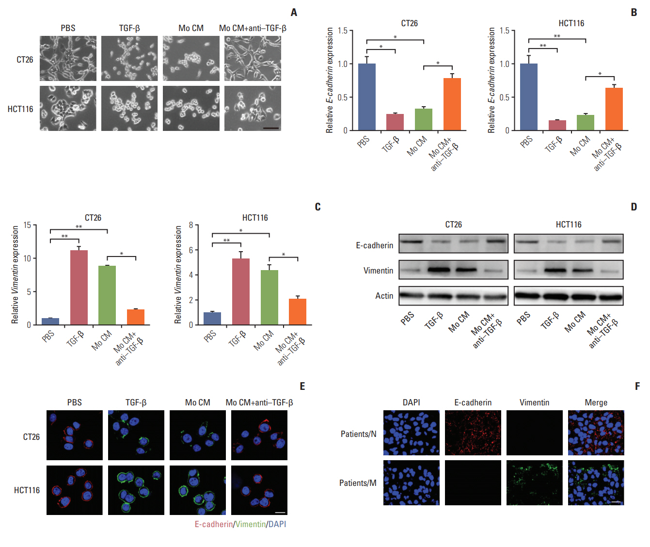
We have found that TGF-β could induce colorectal cancer cells invasion and migration and it has been verified that TGF-β could initiate the EMT to enhance tumor cell invasion and migration in a variety of cancers, which was characterized as loss of E-cadherin and elevated vimentin expression [22-24]. We were wondering that whether TGF-β could trigger EMT in colorectal cancer cells. In order to visually capture the morphological change of EMT in vitro, we conducted the cell imaging of CT26 and HCT116 cells with TGF-β or conditioned medium from TAMs treatment. We observed that treating with TGF-β and conditioned medium from TAMs could significantly induce epithelioid CT26 and HCT-116 cells transition into mesenchymal phenotype (Fig. 3A), while adding anti–TGF-β antibody reversed the effect (Fig. 3A). Moreover, E-cadherin, a hallmark of epithelial cells, was significantly decreased with TGF-β and conditioned medium from TAMs treatment in CT26 and HCT116 cells (Fig. 3B, D, and E). Concomitantly, vimentin, one of the mesenchymal cytoskeletal proteins, was remarkably increased with TGF-β and conditioned medium from TAMs treatment (Fig. 3C-E). Conversely, adding anti‒TGF-β antibody effectively reversed the effect (Fig. 3B-E). These data suggest that TGF-β could induce the colorectal cancer cells EMT. Accumulating evidence supported that EMT was correlated with the metastasis and poor prognosis in cancer patients [25,26]. Here, to confirm colorectal cancer cells metastasis was EMT involved, we analyzed the expression of EMT markers in cancer patients’ tissues. We found that vimentin mainly expressed in metastatic patients’ tissue, while non-metastatic patients’ tissue had plenty E-cadherin expression (Fig. 3F). This clinical data demonstrated that EMT was involved in colorectal cancer cells metastasis. Taking together, those results conclude that TGF-β induced EMT is involved in colorectal cancer cells metastasis.
4. TGF-β mediates colorectal cancer cells EMT via Smad2, 3-4/Snail/E-cadherin pathway
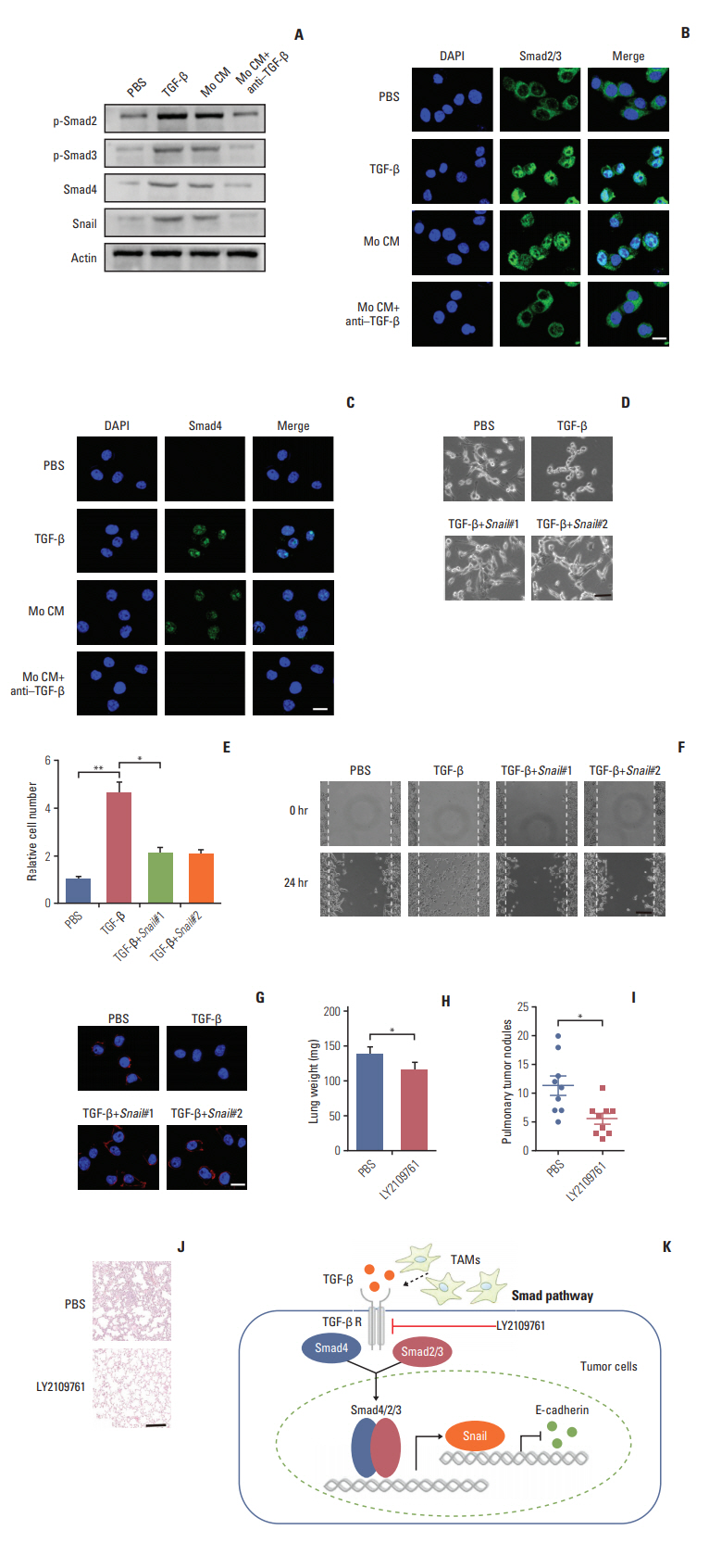
Next, we want to figure out how TGF-β endowed the EMT of colorectal cancer cells and to explore potential therapeutic target in clinic. The signaling mechanisms of TGF-β induced EMT has been largely conducted in a variety of cancer cells. In response to TGF-β, the cellular Smad signaling would be activated by phosphorylated TGF-β receptors [27]. To test whether the Smad signaling involved in the colorectal cancer cells EMT, we detected the activation of Smads with TGF-β or conditioned medium from TAMs in CT26 colorectal cancer cells. We found that TGF-β or conditioned medium from TAMs increased the phosphorylation of Smad2/3, and significantly enhanced the expression of Smad4 (Fig. 4A). Also, we observed that the Smad2/3 and Smad4 complex was translocated into nucleus (Fig. 4B and C). Moreover, Snail, the downstream transcription factor of Smad, was increased by TGF-β and conditioned medium from TAMs treatment (Fig. 4A). While pre-treatment with TGF-β neutralizing antibody, reversed the effect (Fig. 4A-C). Further, siRNA targeting Snail transcripts suppressed the epithelia to mesenchymal phenotype changes in TGF-β treated CT26 cells (Fig. 4D). Furthermore, TGF-β induced colorectal cancer cells invasion and migration were significantly inhibited with Snail knockdown (Fig. 4E and F). In line with the pervious results, TFG-β suppressed the expression of E-cadherin. Here, the silence of Snail could efficiently reverse the effects (Fig. 4G). Those data suggest that TGF-β endowed colorectal cancer cells EMT was Snail dependent. In order to disrupt the TAMs triggered colorectal cancer cells metastasis in vivo, we used TGF-β receptor inhibitor to treat CT26-bearing mice. Exhilaratingly, the lung metastasis was remarkably suppressed in LY21-09761 group compared with PBS group with lighter lung weight and less pulmonary tumor nodules (Fig. 4H-J). Collectively, these results indicate that TGF-β activates the Smads/Snail signaling pathway to induce colorectal cancer cells EMT for metastasis.
Discussion
Cumulated evidences support that TAMs involved in tumor cell metastasis, which is mediated through the secretion of cytokines [28]. Recently, TAMs have been reported to accumulate in the tumor sites and promoted distant metastasis in various cancers [28]. Further studies are required to reveal the mechanism of TAMs and determine whether could improve the clinical efficiency by blocking pathway.
In our study, we found that TAMs isolated from primary tumor tissues promote colorectal cancer cells invasion and migration in vitro and in vivo which provided evidence to support the concept that TAMs play a crucial role in colorectal cancer metastasis. It was consistent with previous research revealing that accumulated TAMs in tumor microenvironment participate in different tumor progression, including cancer stemness maintain, tumor growth and drug resistance, through the secretion of cytokines. Tumor distant metastasis is considered as the main cause of cancer-related deaths, and EMT is regarded as the key steps. Further, our study indicated that TAMs could facilitate the colorectal cancer metastasis through inducing EMT of tumor cells by secretion of TGF-βwhich was validated by in vitro and in vivo data [29]. Also, our results showed that TGF-β produced by TAMs in tumor microenvironments activated the Smad signaling pathway by binding to the TGF-β receptors, followed by the phosphorylated Smad2/Smad3 to form complex with Smad4 and regulate transcription of Snail. Once the TGF-β triggered the colorectal cancer cells EMT, Snail could repress the expression of epithelial marker E-cadherin, resulting in the cancer metastasis (Fig. 4K).
Various crucial features are believed to participate in the cancer metastasis progress, including the expression of integrin [30], tumor cells EMT progression, tumor microenvironments [11]. It has been demonstrated that TAMs play a crucial role in different stages of tumor progression [31]. TAMs facilitate the sustained growth of tumor cells through secretion of growth factors such as CCL8 [20] and remodeling the tumor microenvironments by matrix metalloproteinases (MMPs). Moreover, TAMs promote tumor cells invasion and migration through the secretion of tumor necrosis factor and MMPs [19]. However, the specific mechanism of metastasis induced by TAMs is still unclear. In our studies, we further demonstrate that TAMs could facilitate the metastasis through the secretion of TFG-β in colorectal cancer cells.
Increasing evidences demonstrated that tumor cells could promote metastasis through an autocrine manner. Various cancer cells, including breast cancer cells and pancreatic cancer cells, undergo the progression of EMT before metastasis [29], which is induced by the TGF-β produced by the tumor cells. Our data found that TGF-β could be derived from TAMs and suggested that inhibition of TGF-β signaling significantly decreased the EMT. And the specific signaling pathways of EMT in several cancer cells remained to be unclear. We investigated the progression of EMT in colorectal cancer and found that a Smad dependent signaling pathway, which is induced by the TGF-β produced from TAMs in tumor microenvironment. The data supports TAMs endowed with the capacity to facilitate the metastasis and regulate the tumor progression, readjusting the focus of inhibit tumor growth to selectively suppress TAMs functions and remodeling of tumor microenvironment should be considered when targeting colorectal cancer metastasis in clinic.
In summary, our data provided evidence to support that accumulated TAMs in colorectal cancers contributed to distant metastasis through secreting TGF-βwhich induced EMT by activating Smad2,3-4/Snail pathway. Blocking TGF-β signaling remarkably reduced the EMT which in turn resulted in decreased metastasis. Our data laid an important foundation for potential application of TGF-β inhibition in clinical treatment as anti-metastatic therapy for colorectal cancer patients.
Electronic Supplementary Material
Supplementary materials are available at Cancer Research and Treatment website (https://www.e-crt.org).
Conflict of interest relevant to this article was not reported.
Fig. 1.
Tumor-associated macrophages (TAMs) facilitate colorectal cancer cells metastasis. (A) Quantification of macrophages in tumor tissues of non-metastatic colorectal cancer patients (patients/N) (n=10) and metastatic colorectal cancer patients (patients/M) (n=10). (B) Immunohistochemistry and intensity analysis (n=10) for CD68 to detect the macrophages in tumor tissues of non-metastatic colorectal cancer patients (patients/N) and metastatic colorectal cancer patients (patients/M). Scale bar=100 μm. (C) The lung weight of tumor-bearing mice treated with phosphate buffered saline (PBS) or TAMs (n=10). (D) The lung node number of tumor-bearing mice treated with PBS or TAMs (n=10). (E) H&E staining of a representative lung of tumor-bearing mice treated with PBS or TAMs. Scale bar=200 μm. (F) The lung weight of tumor-bearing mice treated with PBS or clodronate liposomes to deplete macrophages (n=10). (G) The pulmonary tumor nodules of tumor-bearing mice treated with PBS or clodronate liposomes (n=10). (H) H&E staining of a representative lung of tumor-bearing mice treated with PBS or clodronate liposomes. Scale bar=200 μm. The data was presented as the mean±standard error of mean from three independent experiments. *p < 0.05.
Fig. 2.
Tumor-associated macrophages (TAMs) promote colorectal cancer cells metastasis via secretion of transforming growth factor-β (TGF-β). (A) Relative migrant cell number in 104 CT26 and HCT116 cells treated with phosphate buffered saline (PBS) or TAMs cultural supernatant for 24 hours (n=3). (B) The wound-healing assay for evaluating the migration of CT26 and HCT116 cells treated with PBS or TAMs cultural supernatant for 24 hours. Scale bars=100 μm. (C) The relative expression of TGF-β, interleukin (IL)-10, vascular endothelial growth factor (VEGF), CCL18, CCL21, and CXCL21 in peritoneal macrophages or TAMs from tumor tissues of mice (n=3). (D) The relative expression of TGF-βR, IL-10R, vascular endothelial growth factor receptor (VEGFR), PITPNM3, CCR7, and CXCR4 in CT26 treated with TAMs or not (n=3). (E) The content of TGF-β in tumor tissues from non-metastatic or metastatic patients was detected by enzyme-linked immunosorbent assay (n=10). (F) Immunohistochemistry for TGF-β in tumor tissues of non-metastatic colorectal cancer patients (patients/N) and metastatic colorectal cancer patients (patients/M). Scale bar=100 μm. (G) The peritoneal macrophages or TAMs from tumor tissues of mice were isolated to analyze the expression of TGF-β by flow cytometry (n=3). (H) Relative migrant cell number in 104 CT26 cells (n=3) treated with PBS, IL-10, TGF-β, VEGF, CCL18, CCL21, and CXCL12 for 24 hours. (I) The lung weight of tumor-bearing mice treated with PBS, TGFβ, TAMs cultural supernatant, and TAMs cultural supernatant combined with TGF-β neutralizing antibody (n=10). (J) The pulmonary tumor nodules of tumor-bearing mice treated with PBS, TGF-β, TAMs cultural supernatant, and TAMs cultural supernatant combined with TGF-β neutralizing antibody (n=10). (K) H&E staining of a representative lung of tumor-bearing mice treated with PBS, TGF-β, TAMs cultural supernatant, and TAMs cultural supernatant combined with TGF-β neutralizing antibody. Scale bar=200 μm. The data was presented as the mean±SEM from three independent experiments. *p < 0.05, **p < 0.01; ns, not statistically significant.
Fig. 3.
Transforming growth factor-β (TGF-β) induces the colorectal cancer cells metastasis through the epithelial to mesenchymal transition progress. (A) Imagesof CT26 and HCT116 cells treated with phosphate buffered saline (PBS), TGF-β (5 ng/mL), tumor-associated macrophages (TAMs) cultural supernatant and TAMs cultural supernatant combined with TGF-β neutralizing antibody (10 ng/mL) for 24 hours. Scale bar=30 μm. (B) Relative E-cadherin expression of CT26 and HCT116 cells treated with PBS, TGF-β (5 ng/mL), TAMs cultural supernatant, and TAMs cultural supernatant combined with TGF-β neutralizing antibody (10 ng/mL) for 24 hours (n=3). (C) Relative vimentin expression of CT26 and HCT116 cells treated with PBS, TGF-β (5 ng/mL), TAMs cultural supernatant, and TAMs cultural supernatant combined with TGF-β neutralizing antibody (10 ng/mL) for 24 hours (n=3). (D) Western blotting of E-cadherin and vimentin in CT26 and HCT116 cells treated with PBS, TGF-β (5 ng/mL), TAMs cultural supernatant, and TAMs cultural supernatant combined with TGF-β neutralizing antibody (10 ng/mL) for24 hours (E) Immunofluorescence of E-cadherin and vimentin in CT26 and HCT116 cells treated with PBS, TGF-β (5 ng/mL), TAMs cultural supernatant, and TAMs cultural supernatant combined with TGF-β neutralizing antibody (10 ng/mL). Scale bar=10 μm. (F) Immunofluorescence of E-cadherin andvimentin in tumor tissues of non-metastatic colorectal cancer patients (patients/N) andmetastatic colorectal cancer patients (patients/M). Scalebar=20 μm. The data was presented as the mean±standard error of mean from three independent experiments. *p< 0.05, **p< 0.01.
Fig. 4.
Transforming growth factor-β (TGF-β) mediates the colorectal cancer cells epithelial to mesenchymal transition progress via Smad2,3-4/Snail/E-cadherin pathway. (A) Western blotting of p-Smad2, p-Smad3, Smad4, Snail in CT26 cells treated with phosphate buffered saline (PBS), TGF-β (5 ng/mL), tumor-associated macrophages (TAMs) cultural supernatant, and TAMs cultural supernatant combined with TGF-β neutralizing antibody (10 ng/mL). (B) Immunofluorescence of Smad2/3 in CT26 cells treated with PBS, TGF-β (5 ng/mL), TAMs cultural supernatant, and TAMs cultural supernatant combined with TGF-β neutralizing antibody (10 ng/mL). Scale bar=10 μm. (C) Immunofluorescence of Smad4 in CT26 cells treated with PBS, TGF-β (5 ng/mL), TAMs cultural supernatant, and TAMs cultural supernatant combined with TGF-β neutralizing antibody (10 ng/mL). Scale bar=10 μm. (D) Images of CT26 cells treated with PBS, TGF-β, and TGF-β combined with Snail siRNA. Scale bar=20 μm. (E) Relative migrant cell number in 104 CT26 cells treated with PBS, TGF-β, and TGF-β combined with Snail siRNA (n=3). (F) The wound-healing assay for evaluating the migration of CT26 treated with PBS, TGF-β, and TGF-β combined with Snail siRNA. Scale bar=100 μm. (G) Immunofluorescence of E-cadherin in CT26
treated with PBS, TGF-β, and TGF-β combined with Snail siRNA. Scale bar=10 μm. (H) The lung weight of tumor-bearing mice treated with PBS or LY2109761 (n=9). (I) The pulmonary tumor nodules of tumor-bearing mice treated with PBS or LY2109761 (n=9). (J) H&E staining of a representative lung of tumor-bearing mice treated with PBS or LY2109761. Scale bar=200 μm. (K) Schema of TAMs inducing the colorectal cancer cells metastasis. The data was presented as the mean±standard error of mean from three independent experiments. *p < 0.05, **p < 0.01.
Table 1.
The clinical characteristics of patients used in the paper
No. (%) (n=40)
Age (yr)
≥ 60
24 (60.0)
< 60
16 (40.0)
Sex
Female
11 (27.5)
Male
29 (72.5)
Tumor size (mm)
≥ 40 (large)
15 (27.5)
< 40 (small)
25 (62.5)
Metastasis
Absent
20 (50.0)
Present
20 (50.0)
REFERENCES
1. Siegel RL, Miller KD, Jemal A. Cancer statistics, 2017. CA Cancer J Clin. 2017;67:7–30. ArticlePubMed
2. Kim IH, Lee JE, Yang JH, Jeong JW, Ro S, Oh ST, et al. Clinical significance of discordance between carcinoembryonic antigen levels and RECIST in metastatic colorectal cancer. Cancer Res Treat. 2018;50:283–92. ArticlePubMedPDF
3. Loupakis F, Cremolini C, Masi G, Lonardi S, Zagonel V, Salvatore L, et al. Initial therapy with FOLFOXIRI and bevacizumab for metastatic colorectal cancer. N Engl J Med. 2014;371:1609–18. ArticlePubMed
4. O'Connell JB, Maggard MA, Ko CY. Colon cancer survival rates with the new American Joint Committee on Cancer sixth edition staging. J Natl Cancer Inst. 2004;96:1420–5. ArticlePubMed
5. Simmonds PC, Primrose JN, Colquitt JL, Garden OJ, Poston GJ, Rees M. Surgical resection of hepatic metastases from colorectal cancer: a systematic review of published studies. Br J Cancer. 2006;94:982–99. ArticlePubMedPMCPDF
6. Liu H, Radisky DC, Yang D, Xu R, Radisky ES, Bissell MJ, et al. MYC suppresses cancer metastasis by direct transcriptional silencing of alphav and beta3 integrin subunits. Nat Cell Biol. 2012;14:567–74. ArticlePubMedPMCPDF
7. Ma L, Young J, Prabhala H, Pan E, Mestdagh P, Muth D, et al. miR-9, a MYC/MYCN-activated microRNA, regulates E-cadherin and cancer metastasis. Nat Cell Biol. 2010;12:247–56. ArticlePubMedPMCPDF
8. Tanaka H, Kono E, Tran CP, Miyazaki H, Yamashiro J, Shimomura T, et al. Monoclonal antibody targeting of N-cadherin inhibits prostate cancer growth, metastasis and castration resistance. Nat Med. 2010;16:1414–20. ArticlePubMedPMCPDF
9. Yoo YA, Kang MH, Lee HJ, Kim BH, Park JK, Kim HK, et al. Sonic hedgehog pathway promotes metastasis and lymphangiogenesis via activation of Akt, EMT, and MMP-9 pathway in gastric cancer. Cancer Res. 2011;71:7061–70. ArticlePubMed
10. Gilkes DM, Semenza GL, Wirtz D. Hypoxia and the extracellular matrix: drivers of tumour metastasis. Nat Rev Cancer. 2014;14:430–9. ArticlePubMedPMCPDF
11. Kessenbrock K, Plaks V, Werb Z. Matrix metalloproteinases: regulators of the tumor microenvironment. Cell. 2010;141:52–67. ArticlePubMedPMC
12. Chaffer CL, Weinberg RA. A perspective on cancer cell metastasis. Science. 2011;331:1559–64. ArticlePubMed
13. Ye X, Brabletz T, Kang Y, Longmore GD, Nieto MA, Stanger BZ, et al. Upholding a role for EMT in breast cancer metastasis. Nature. 2017;547:E1–3. ArticlePubMedPMC
14. Steidl C, Lee T, Shah SP, Farinha P, Han G, Nayar T, et al. Tumor-associated macrophages and survival in classic Hodgkin's lymphoma. N Engl J Med. 2010;362:875–85. ArticlePubMedPMC
15. Franklin RA, Liao W, Sarkar A, Kim MV, Bivona MR, Liu K, et al. The cellular and molecular origin of tumor-associated macrophages. Science. 2014;344:921–5. ArticlePubMedPMC
16. Ye XZ, Xu SL, Xin YH, Yu SC, Ping YF, Chen L, et al. Tumorassociated microglia/macrophages enhance the invasion of glioma stem-like cells via TGF-beta1 signaling pathway. J Immunol. 2012;189:444–53. ArticlePubMed
17. Erreni M, Mantovani A, Allavena P. Tumor-associated macrophages (TAM) and inflammation in colorectal cancer. Cancer Microenviron. 2011;4:141–54. ArticlePubMed
18. Ikushima H, Miyazono K. TGFbeta signalling: a complex web in cancer progression. Nat Rev Cancer. 2010;10:415–24. ArticlePubMedPDF
19. Chaudhury A, Hussey GS, Ray PS, Jin G, Fox PL, Howe PH. TGF-beta-mediated phosphorylation of hnRNP E1 induces EMT via transcript-selective translational induction of Dab2 and ILEI. Nat Cell Biol. 2010;12:286–93. ArticlePubMedPMCPDF
20. Chen J, Yao Y, Gong C, Yu F, Su S, Chen J, et al. CCL18 from tumor-associated macrophages promotes breast cancer metastasis via PITPNM3. Cancer Cell. 2011;19:541–55. ArticlePubMedPMC
21. Sica A, Schioppa T, Mantovani A, Allavena P. Tumour-associated macrophages are a distinct M2 polarised population promoting tumour progression: potential targets of anti-cancer therapy. Eur J Cancer. 2006;42:717–27. ArticlePubMed
22. Braun J, Hoang-Vu C, Dralle H, Huttelmaier S. Downregulation of microRNAs directs the EMT and invasive potential of anaplastic thyroid carcinomas. Oncogene. 2010;29:4237–44. ArticlePubMedPDF
23. Zhang H, Liu L, Wang Y, Zhao G, Xie R, Liu C, et al. KLF8 involves in TGF-beta-induced EMT and promotes invasion and migration in gastric cancer cells. J Cancer Res Clin Oncol. 2013;139:1033–42. ArticlePubMed
24. Padua D, Massague J. Roles of TGFbeta in metastasis. Cell Res. 2009;19:89–102. ArticlePubMedPDF
25. Kudo-Saito C, Shirako H, Takeuchi T, Kawakami Y. Cancer metastasis is accelerated through immunosuppression during Snail-induced EMT of cancer cells. Cancer Cell. 2009;15:195–206. ArticlePubMed
26. Mulholland DJ, Kobayashi N, Ruscetti M, Zhi A, Tran LM, Huang J, et al. Pten loss and RAS/MAPK activation cooperate to promote EMT and metastasis initiated from prostate cancer stem/progenitor cells. Cancer Res. 2012;72:1878–89. ArticlePubMedPMC
27. Feng XH, Derynck R. Specificity and versatility in tgf-beta signaling through Smads. Annu Rev Cell Dev Biol. 2005;21:659–93. ArticlePubMed
28. Qian BZ. Inflammation fires up cancer metastasis. Semin Cancer Biol. 2017;47:170–6. ArticlePubMed
29. David CJ, Huang YH, Chen M, Su J, Zou Y, Bardeesy N, et al. TGF-beta tumor suppression through a lethal EMT. Cell. 2016;164:1015–30. ArticlePubMedPMC
30. Seguin L, Desgrosellier JS, Weis SM, Cheresh DA. Integrins and cancer: regulators of cancer stemness, metastasis, and drug resistance. Trends Cell Biol. 2015;25:234–40. ArticlePubMedPMC
The role of matrix metalloproteinase-2 in the metastatic cascade: a review Nigel P. Murray Oncologie.2024; 26(1): 27. CrossRef
Tumor-associated macrophages in colorectal cancer metastasis: molecular insights and translational perspectives Siyu Hou, Yuanchun Zhao, Jiajia Chen, Yuxin Lin, Xin Qi Journal of Translational Medicine.2024;[Epub] CrossRef
The role of stromal cells in epithelial–mesenchymal plasticity and its therapeutic potential Juanjing Wang, Junmei Peng, Yonglin Chen, M. I. Nasser, Hui Qin Discover Oncology.2024;[Epub] CrossRef
Isolation and purification of polysaccharides from Bupleurum marginatum Wall.ex DC and their anti-liver fibrosis activities Li Xiao, Hafsa Sunniya, Jingyi Li, Mohib Ullah Kakar, Rongji Dai, Bo Li Frontiers in Pharmacology.2024;[Epub] CrossRef
Key players of immunosuppression in epithelial malignancies: Tumor‐infiltrating myeloid cells and γδ T cells Baishali Tamuli, Sakshi Sharma, Meena Patkar, Subir Biswas Cancer Reports.2024;[Epub] CrossRef
Correlation of tumor-associated macrophage density and proportion of M2 subtypes with the pathological stage of colorectal cancer Fouzia Fazal, Muhammad Arsalan Khan, Sumayya Shawana, Rahma Rashid, Muhammed Mubarak World Journal of Gastrointestinal Oncology.2024; 16(5): 1878. CrossRef
Reprogramming Tumor-Associated Macrophage Using Nanocarriers: New Perspectives to Halt Cancer Progression Alyona B. Kuznetsova, Ekaterina P. Kolesova, Alessandro Parodi, Andrey A. Zamyatnin, Vera S. Egorova Pharmaceutics.2024; 16(5): 636. CrossRef
Deciphering the performance of macrophages in tumour microenvironment: a call for precision immunotherapy Belén Toledo, Linrui Zhu Chen, María Paniagua-Sancho, Juan Antonio Marchal, Macarena Perán, Elisa Giovannetti Journal of Hematology & Oncology.2024;[Epub] CrossRef
Unveiling the contribution of tumor-associated macrophages in driving epithelial-mesenchymal transition: a review of mechanisms and therapeutic Strategies Yijia Zhang, Xiaofei Ding, Xue Zhang, Ye Li, Rui Xu, Hai-Jun Li, Daiying Zuo, Guang Chen Frontiers in Pharmacology.2024;[Epub] CrossRef
The Role of Adipocytes Recruited as Part of Tumor Microenvironment in Promoting Colorectal Cancer Metastases Yunxia Ma, Miljana Nenkov, Yuan Chen, Nikolaus Gaßler International Journal of Molecular Sciences.2024; 25(15): 8352. CrossRef
Polarization of M2 Tumor-Associated Macrophages (TAMs) in Cancer Immunotherapy
Indy Bui, Benjamin Bonavida Critical Reviews™ in Oncogenesis.2024; 29(4): 75. CrossRef
Mapping cellular interactions from spatially resolved transcriptomics data James Zhu, Yunguan Wang, Woo Yong Chang, Alicia Malewska, Fabiana Napolitano, Jeffrey C. Gahan, Nisha Unni, Min Zhao, Rongqing Yuan, Fangjiang Wu, Lauren Yue, Lei Guo, Zhuo Zhao, Danny Z. Chen, Raquibul Hannan, Siyuan Zhang, Guanghua Xiao, Ping Mu, Ariell Nature Methods.2024; 21(10): 1830. CrossRef
Elevated BEAN1 expression correlates with poor prognosis, immune evasion, and chemotherapy resistance in rectal adenocarcinoma Tiannake Shapaer, Yi Chen, Yipeng Pan, Zhimin Wu, Tuoxian Tang, Zeliang Zhao, Xiangyue Zeng Discover Oncology.2024;[Epub] CrossRef
MicroRNA-382 as a tumor suppressor during tumor progression Yalda Samsami, Iman Akhlaghipour, Negin Taghehchian, Mahsa Palizkaran Yazdi, Saba Farrokhi, Hamid Reza Rahimi, Meysam Moghbeli Bioorganic & Medicinal Chemistry Letters.2024; 113: 129967. CrossRef
Research progress of tumor-associated macrophages in immune checkpoint inhibitor tolerance in colorectal cancer Qi Fan, Zheng-Wei Fu, Ming Xu, Feng Lv, Jia-Song Shi, Qi-Qi Zeng, De-Hai Xiong World Journal of Gastrointestinal Oncology.2024; 16(10): 4064. CrossRef
Lidocaine combined with general anesthetics impedes metastasis of breast cancer cells via inhibition of TGF-β/Smad-mediated EMT signaling by reprogramming tumor-associated macrophages Beom Seok Han, Soyeon Ko, Min Seok Park, Yun Ji Lee, Sang Eun Kim, Pureunchowon Lee, Ye Jin Cho, Han Gyeol Go, Sehan Kwak, Eunji Park, Ayoung Lim, Suji Lee, Seungjong Yoo, Hyunzu Kim, Kyung Hee Jung, Soon-Sun Hong International Immunopharmacology.2024; 142: 113207. CrossRef
Targeted nanomedicine for reprogramming the tumor innate immune system: From bench to bedside Kunal Pednekar, Julia Minnee, I. Jolanda M. de Vries, Jai Prakash European Journal of Pharmaceutics and Biopharmaceutics.2024; 204: 114510. CrossRef
Natural compounds modulate the mechanism of action of tumour-associated macrophages against colorectal cancer: a review Weichen Yuan, Jiexiang Zhang, Haibin Chen, Yupei Zhuang, Hongli Zhou, Wenting Li, Wenli Qiu, Hongguang Zhou Journal of Cancer Research and Clinical Oncology.2024;[Epub] CrossRef
Retinoic acid-loaded PLGA nanocarriers targeting cell cholesterol potentialize the antitumour effect of PD-L1 antibody by preventing epithelial-mesenchymal transition mediated by M2-TAM in colorectal cancer Raimundo Fernandes de Araújo Júnior, George A Lira, Timo Schomann, Rômulo S Cavalcante, Natalia Feitosa Vilar, Regina Célia Monteiro de Paula, Raelle Ferreira Gomes, Chih Kit Chung, Carla Jorquera-Cordero, Olena Vepris, Alan B Chan, Luis J. Cruz Translational Oncology.2023; 31: 101647. CrossRef
The ceRNA network regulates epithelial-mesenchymal transition in colorectal cancer Ruina Li, Hui Xu, Xiaoling Gao Heliyon.2023; 9(3): e14143. CrossRef
Do Elevated YKL-40 Levels Drive the Immunosuppressive Tumor Microenvironment in Colorectal Cancer? Assessment of the Association of the Expression of YKL-40, MMP-8, IL17A, and PD-L1 with Coexisting Type 2 Diabetes, Obesity, and Active Smoking Błażej Ochman, Sylwia Mielcarska, Agnieszka Kula, Miriam Dawidowicz, Julia Robotycka, Jerzy Piecuch, Monika Szrot, Sylwia Dzięgielewska-Gęsiak, Małgorzata Muc-Wierzgoń, Dariusz Waniczek, Elżbieta Świętochowska Current Issues in Molecular Biology.2023; 45(4): 2781. CrossRef
TGFβ and the Tumor Microenvironment in Colorectal Cancer Maximilian J. Waldner, Markus F. Neurath Cells.2023; 12(8): 1139. CrossRef
Exosomes in metastasis of colorectal cancers: Friends or foes? Zheng Wu, Ze-Xuan Fang, Yan-Yu Hou, Bing-Xuan Wu, Yu Deng, Hua-Tao Wu, Jing Liu World Journal of Gastrointestinal Oncology.2023; 15(5): 731. CrossRef
Dynamic polarization of tumor-associated macrophages and their interaction with intratumoral T cells in an inflamed tumor microenvironment: from mechanistic insights to therapeutic opportunities Jiashu Han, Luochu Dong, Mengwei Wu, Fei Ma Frontiers in Immunology.2023;[Epub] CrossRef
MicroRNA functional metal-organic framework nanocomposite for efficient inhibition of drug-resistant breast cancer cells Yuxin Shen, Yao Zhang, Xiyue Gao, Mengdi Shang, Yanfei Cai, Zhaoqi Yang Emergent Materials.2023; 6(5): 1537. CrossRef
Impact of Opioids on Cellular Metabolism: Implications for Metabolic Pathways Involved in Cancer Doorsa Tarazi, Jason T. Maynes Pharmaceutics.2023; 15(9): 2225. CrossRef
Suppressing epithelial‐mesenchymal‐transition blue light therapy for reducing macrophage‐mediated cancerous pulmonary fibrosis: An in‐vitro study Wenjun Zhang, Jianfei Dong Journal of Biophotonics.2023;[Epub] CrossRef
Metastasis organotropism in colorectal cancer: advancing toward innovative therapies Kai He, Zhihan Wang, Maochao Luo, Bowen Li, Ning Ding, Lei Li, Bo He, Han Wang, Jiangjun Cao, Canhua Huang, Jun Yang, Hai-Ning Chen Journal of Translational Medicine.2023;[Epub] CrossRef
Thermal/ultrasound-triggered release of liposomes loaded with Ganoderma applanatum polysaccharide from microbubbles for enhanced tumour ablation Jiani Yuan, Lei Ding, Lu Han, Lina Pang, Peidi Zhang, Xiao Yang, Haijing Liu, Minjuan Zheng, Yunfei Zhang, Wen Luo Journal of Controlled Release.2023; 363: 84. CrossRef
MicroRNAs and their vital role in apoptosis in hepatocellular carcinoma: miRNA-based diagnostic and treatment methods Masoumeh Hajizadeh, Farnaz Hajizadeh, Sevil Ghaffarei, Mohammad Amin Doustvandi, Khadijeh Hajizadeh, Seyyed Mohammad Yaghoubi, Fateme Mohammadnejad, Nadia Allahyarzadeh Khiabani, Pegah Mousavi, Behzad Baradaran Gene.2023; 888: 147803. CrossRef
Tumour-associated macrophages: versatile players in the tumour microenvironment Zoey Zeyuan Ji, Max Kam-Kwan Chan, Alex Siu-Wing Chan, Kam-Tong Leung, Xiaohua Jiang, Ka-Fai To, Yi Wu, Patrick Ming-Kuen Tang Frontiers in Cell and Developmental Biology.2023;[Epub] CrossRef
Exploiting innate immunity for cancer immunotherapy Ming Yi, Tianye Li, Mengke Niu, Qi Mei, Bin Zhao, Qian Chu, Zhijun Dai, Kongming Wu Molecular Cancer.2023;[Epub] CrossRef
Functional and Therapeutic Significance of Tumor-Associated Macrophages in Colorectal Cancer Yitong Li, Zhenmei Chen, Jiahao Han, Xiaochen Ma, Xin Zheng, Jinhong Chen Frontiers in Oncology.2022;[Epub] CrossRef
Targeting Epithelial-to-Mesenchymal Transition in Radioresistance: Crosslinked Mechanisms and Strategies Lili Qiao, Yanfei Chen, Ning Liang, Jian Xie, Guodong Deng, Fangjie Chen, Xiaojuan Wang, Fengjun Liu, Yupeng Li, Jiandong Zhang Frontiers in Oncology.2022;[Epub] CrossRef
SNAIL driven by a feed forward loop motif promotes TGFβ induced epithelial to mesenchymal transition Gottumukkala Sai Bhavani, Anbumathi Palanisamy Biomedical Physics & Engineering Express.2022; 8(4): 045012. CrossRef
Comparative analysis of tumor control gene and microRNA expression profiles in tumor and adjacent tissues in patients with colorectal cancer M. I. Sluzhev, M. I. Zaraisky, V. V. Semiglazov, T. Yu. Semiglazova, E. V. Tkachenko, S. V. Kondratev, N. A. Brish, Yu. V. Alekseeva, Iu. V. Petrik, A. N. Sidorova Siberian journal of oncology.2022; 21(2): 55. CrossRef
D-tryptophan triggered epithelial-mesenchymal transition by activating TGF-β signaling pathway Chong Wang, Fangting Wang, Yanbo Wang, Linglin Fu Food Science and Human Wellness.2022; 11(5): 1215. CrossRef
AST·MLR index and operation injury condition are novel prognostic predictor for the prediction of survival in patients with colorectal cancer liver metastases undergoing surgical resection Qichen Chen, Mingxia Li, Jinghua Chen, Zhen Huang, Xiao Chen, Hong Zhao, Jianqiang Cai BMC Cancer.2022;[Epub] CrossRef
Molecular Characterizations of Gynecologic Carcinosarcomas: A Focus on the Immune Microenvironment Sanaa Nakad Borrego, Ernst Lengyel, Katherine C. Kurnit Cancers.2022; 14(18): 4465. CrossRef
miR‑382 inhibits breast cancer progression and metastasis by affecting the M2 polarization of tumor‑associated macrophages by targeting PGC‑1α Hua Zhou, Mingyu Gan, Xin Jin, Meng Dai, Yuanyuan Wang, Youyang Lei, Zijing Lin, Jia Ming International Journal of Oncology.2022;[Epub] CrossRef
TGF-β Signaling in Metastatic Colorectal Cancer (mCRC): From Underlying Mechanism to Potential Applications in Clinical Development Xiaoshuang Li, Yanmin Wu, Tian Tian International Journal of Molecular Sciences.2022; 23(22): 14436. CrossRef
Naringenin in the Prevention of Colon Cancer: An Updated Review
Vemula Sarojamma, Ramakrishna Vadde Onco Therapeutics.2022; 9(2): 25. CrossRef
Relationship between Epithelial-to-Mesenchymal Transition and Tumor-Associated Macrophages in Colorectal Liver Metastases Aurora Gazzillo, Michela Anna Polidoro, Cristiana Soldani, Barbara Franceschini, Ana Lleo, Matteo Donadon International Journal of Molecular Sciences.2022; 23(24): 16197. CrossRef
CD147 deficiency in T cells prevents thymic involution by inhibiting the EMT process in TECs in the presence of TGFβ Ruo Chen, Ke Wang, Zhuan Feng, Ming-Yang Zhang, Jiao Wu, Jie-Jie Geng, Zhi-Nan Chen Cellular & Molecular Immunology.2021; 18(1): 171. CrossRef
TGFβ drives NK cell metabolic dysfunction in human metastatic breast cancer Karen Slattery, Elena Woods, Vanessa Zaiatz-Bittencourt, Sam Marks, Sonya Chew, Michael Conroy, Caitriona Goggin, Colm MacEochagain, John Kennedy, Sophie Lucas, David K Finlay, Clair M Gardiner Journal for ImmunoTherapy of Cancer.2021; 9(2): e002044. CrossRef
Long non-coding RNA ZFAS1 is a major regulator of epithelial-mesenchymal transition through miR-200/ZEB1/E-cadherin, vimentin signaling in colon adenocarcinoma Stephen J. O’Brien, Casey Fiechter, James Burton, Jacob Hallion, Mason Paas, Ankur Patel, Ajay Patel, Andre Rochet, Katharina Scheurlen, Sarah Gardner, Maurice Eichenberger, Harshini Sarojini, Sudhir Srivastava, Shesh Rai, Theodore Kalbfleisch, Hiram C. P Cell Death Discovery.2021;[Epub] CrossRef
Creatine promotes cancer metastasis through activation of Smad2/3 Liwen Zhang, Zijing Zhu, Huiwen Yan, Wen Wang, Zhenzhen Wu, Fei Zhang, Qixiang Zhang, Guizhi Shi, Junfeng Du, Huiyun Cai, Xuanxuan Zhang, David Hsu, Pu Gao, Hai-long Piao, Gang Chen, Pengcheng Bu Cell Metabolism.2021; 33(6): 1111. CrossRef
Lactate Dehydrogenase-A (LDH-A) Preserves Cancer Stemness and Recruitment of Tumor-Associated Macrophages to Promote Breast Cancer Progression Shengnan Wang, Lingyu Ma, Ziyuan Wang, Huiwen He, Huilin Chen, Zhaojun Duan, Yuyang Li, Qin Si, Tsung-Hsien Chuang, Chong Chen, Yunping Luo Frontiers in Oncology.2021;[Epub] CrossRef
Partial EMT in head and neck cancer biology: a spectrum instead of a switch Ananya Pal, Thomas F. Barrett, Rachel Paolini, Anuraag Parikh, Sidharth V. Puram Oncogene.2021; 40(32): 5049. CrossRef
Macrophages, as a Promising Strategy to Targeted Treatment for Colorectal Cancer Metastasis in Tumor Immune Microenvironment Yingru Zhang, Yiyang Zhao, Qi Li, Yan Wang Frontiers in Immunology.2021;[Epub] CrossRef
Tumor‐associated macrophage‐derived CCL5 promotes chemotherapy resistance and metastasis in prostatic cancer Jian Ma, Fuerhaiti Shayiti, Jing Ma, Meng Wei, Tingting Hua, Rong Zhang, Junyan Su, Peng Chen Cell Biology International.2021; 45(10): 2054. CrossRef
Tumor Microenvironment: Involved Factors and Signaling Pathways in Epithelial-Mesenchymal Transition Madeh Ghaderi, Azadeh Niknejad International Journal of Cancer Management.2021;[Epub] CrossRef
The Multifaceted Role of TGF-β in Gastrointestinal Tumors Fabio Sabbadini, Monica Bertolini, Serena De Matteis, Domenico Mangiameli, Serena Contarelli, Silvia Pietrobono, Davide Melisi Cancers.2021; 13(16): 3960. CrossRef
TMEM100 Modulates TGF-β Signaling Pathway to Inhibit Colorectal Cancer Progression Huixia Li, Chuan Cheng, Weibo You, Jiujian Zheng, Jie Xu, Peng Gao, Jianping Wang, Tatsuya Toyokawa Gastroenterology Research and Practice.2021; 2021: 1. CrossRef
Green Synthesized Honokiol Transfersomes Relieve the Immunosuppressive and Stem-Like Cell Characteristics of the Aggressive B16F10 Melanoma Yasmeen Ezzeldeen, Shady Swidan, Aliaa ElMeshad, Aya Sebak International Journal of Nanomedicine.2021; Volume 16: 5693. CrossRef
Copper content in ascitic fluid is associated with angiogenesis and progression in ovarian cancer Toshimichi Onuma, Tetsuya Mizutani, Yuko Fujita, Shizuka Yamada, Yoshio Yoshida Journal of Trace Elements in Medicine and Biology.2021; 68: 126865. CrossRef
KLK8 promotes the proliferation and metastasis of colorectal cancer via the activation of EMT associated with PAR1 Qing Hua, Zhirong Sun, Yi Liu, Xuefang Shen, Weiwei Zhao, Xiaoyan Zhu, Pingbo Xu Cell Death & Disease.2021;[Epub] CrossRef
Tumor-Released Products Promote Bone Marrow-Derived Macrophage Survival and Proliferation Juliana Maria Motta, Vivian Mary Rumjanek, Alberto Mantovani, Massimo Locati Biomedicines.2021; 9(10): 1387. CrossRef
Potential role of tumor-infiltrating T-, B-lymphocytes, tumor-associated macrophages and IgA-secreting plasma cells in long-term survival in the rectal adenocarcinoma patients Dmitry A. Zinovkin, Suheyla Y. Kose, Eldar A. Nadyrov, Sergey L. Achinovich, Dmitry M. Los', Tatyana E. Gavrilenko, Dmitry I. Gavrilenko, Jale Yuzugulen, Md Zahidul Islam Pranjol Life Sciences.2021; 286: 120052. CrossRef
Targeting Tumor-Associated Macrophages in Cancer Immunotherapy Amy J. Petty, Dwight H. Owen, Yiping Yang, Xiaopei Huang Cancers.2021; 13(21): 5318. CrossRef
Tumour microenvironment: a non-negligible driver for epithelial−mesenchymal transition in colorectal cancer Lei Han, Shuyi Wang, Chen Wei, Yan Fang, Sihao Huang, Tailang Yin, Bin Xiong, Chaogang Yang Expert Reviews in Molecular Medicine.2021;[Epub] CrossRef
Anti-Cancer Activity Profiling of Chemotherapeutic Agents in 3D Co-Cultures of Pancreatic Tumor Spheroids with Cancer-Associated Fibroblasts and Macrophages So-Dam Jang, Jeeyeun Song, Hyun-Ah Kim, Chang-Nim Im, Iftikhar Ali Khawar, Jong Kook Park, Hyo-Jeong Kuh Cancers.2021; 13(23): 5955. CrossRef
M2-Type Macrophages Induce Tregs Generation by Activating the TGF-β/Smad Signalling Pathway to Promote Colorectal Cancer Development Xueqian Ma, Yu Gao, Yanlei Chen, Jinhao Liu, Chunyu Yang, Cuifen Bao, Yanping Wang, Yang Feng, Xiaoyu Song, Shifeng Qiao OncoTargets and Therapy.2021; Volume 14: 5391. CrossRef
Communication between EMT and PD-L1 signaling: New insights into tumor immune evasion Yuanyuan Jiang, Hanxiang Zhan Cancer Letters.2020; 468: 72. CrossRef
Signaling of Macrophages that Contours the Tumor Microenvironment for Promoting Cancer Development Justin K. Messex, Crystal J. Byrd, Geou-Yarh Liou Cells.2020; 9(4): 919. CrossRef
The roles of tumor-associated macrophages in tumor angiogenesis and metastasis Luo-Qin Fu, Wen-Lin Du, Mao-Hua Cai, Jia-Yu Yao, Yuan-Yuan Zhao, Xiao-Zhou Mou Cellular Immunology.2020; 353: 104119. CrossRef
Pro-tumorigenic functions of macrophages at the primary, invasive and metastatic tumor site Elaheh Nasrollahzadeh, Sepideh Razi, Mahsa Keshavarz-Fathi, Massimiliano Mazzone, Nima Rezaei Cancer Immunology, Immunotherapy.2020; 69(9): 1673. CrossRef
Clinical Implication of Concordant or Discordant Genomic Profiling between Primary and Matched Metastatic Tissues in Patients with Colorectal Cancer Jung Yoon Choi, Sunho Choi, Minhyeok Lee, Young Soo Park, Jae Sook Sung, Won Jin Chang, Ju Won Kim, Yoon Ji Choi, Jin Kim, Dong-Sik Kim, Sung-Ho Lee, Junhee Seok, Kyong Hwa Park, Seon Hahn Kim, Yeul Hong Kim Cancer Research and Treatment.2020; 52(3): 764. CrossRef
Caspase Recruitment Domain Containing Protein 9 Suppresses Non-Small Cell Lung Cancer Proliferation and Invasion via Inhibiting MAPK/p38 Pathway Linyue Pan, Yuting Tan, Bin Wang, Wenjia Qiu, Yulei Yin, Haiyan Ge, Huili Zhu Cancer Research and Treatment.2020; 52(3): 867. CrossRef
RUNX1 regulates TGF-β induced migration and EMT in colorectal cancer Chenghao Lu, Zheyu Yang, Dingye Yu, Jianwei Lin, Wei Cai Pathology - Research and Practice.2020; 216(11): 153142. CrossRef
Targeting Tumor-Associated Macrophages in Anti-Cancer Therapies: Convincing the Traitors to Do the Right Thing Cristina Belgiovine, Elisabeth Digifico, Clément Anfray, Aldo Ummarino, Fernando Torres Andón Journal of Clinical Medicine.2020; 9(10): 3226. CrossRef
Mapping a Circular RNA–microRNA–mRNA-Signaling Regulatory Axis that Modulates Stemness Properties of Cancer Stem Cell Populations in Colorectal Cancer Spheroid Cells Vimalan Rengganaten, Chiu-Jung Huang, Ping-Hsing Tsai, Mong-Lien Wang, Yi-Ping Yang, Yuan-Tzu Lan, Wen-Liang Fang, Shelly Soo, Hooi Tin Ong, Soon Keng Cheong, Kong Bung Choo, Shih-Hwa Chiou International Journal of Molecular Sciences.2020; 21(21): 7864. CrossRef
A Window of Opportunity: Targeting Cancer Endothelium to Enhance Immunotherapy Gizem Duru, Marjolein van Egmond, Niels Heemskerk Frontiers in Immunology.2020;[Epub] CrossRef
Modeling of the immune response in the pathogenesis of solid tumors and its prognostic significance Łukasz Zadka, Damian J. Grybowski, Piotr Dzięgiel Cellular Oncology.2020; 43(4): 539. CrossRef
Overview of the oncogenic signaling pathways in colorectal cancer: Mechanistic insights Ammad Ahmad Farooqi, Marc de la Roche, Mustafa B.A. Djamgoz, Zahid H. Siddik Seminars in Cancer Biology.2019;[Epub] CrossRef
Transcriptome Sequencing Investigated the Tumor-Related Factors Changes After T. gondii Infection Gang Lu, Jian Zhou, Ying hui Zhao, Qiao ling Li, Yun yun Gao, Lin Wang Frontiers in Microbiology.2019;[Epub] CrossRef
Advances in Molecular Mechanisms and Immunotherapy Involving the Immune Cell-Promoted Epithelial-to-Mesenchymal Transition in Lung Cancer Serena De Matteis, Matteo Canale, Alberto Verlicchi, Giuseppe Bronte, Angelo Delmonte, Lucio Crinò, Giovanni Martinelli, Paola Ulivi Journal of Oncology.2019; 2019: 1. CrossRef
Tumor-associated macrophages: an accomplice in solid tumor progression Yibing Chen, Yucen Song, Wei Du, Longlong Gong, Haocai Chang, Zhengzhi Zou Journal of Biomedical Science.2019;[Epub] CrossRef
Transforming Growth Factor-β Signaling Pathway in Colorectal Cancer and Its Tumor Microenvironment Yoshiro Itatani, Kenji Kawada, Yoshiharu Sakai International Journal of Molecular Sciences.2019; 20(23): 5822. CrossRef
Gastric cancer-derived mesenchymal stromal cells trigger M2 macrophage polarization that promotes metastasis and EMT in gastric cancer Wei Li, Xu Zhang, Fenglei Wu, Ying Zhou, Zengtao Bao, Haining Li, Ping Zheng, Shaolin Zhao Cell Death & Disease.2019;[Epub] CrossRef
Pimozide suppresses colorectal cancer via inhibition of Wnt/β-catenin signaling pathway Yonggang Ren, Jia Tao, Zhen Jiang, Dongmei Guo, Jiancai Tang Life Sciences.2018; 209: 267. CrossRef
Tumor-Associated Macrophages Derived TGF-β‒Induced Epithelial to Mesenchymal Transition in Colorectal Cancer Cells through Smad2,3-4/Snail Signaling Pathway
Fig. 1. Tumor-associated macrophages (TAMs) facilitate colorectal cancer cells metastasis. (A) Quantification of macrophages in tumor tissues of non-metastatic colorectal cancer patients (patients/N) (n=10) and metastatic colorectal cancer patients (patients/M) (n=10). (B) Immunohistochemistry and intensity analysis (n=10) for CD68 to detect the macrophages in tumor tissues of non-metastatic colorectal cancer patients (patients/N) and metastatic colorectal cancer patients (patients/M). Scale bar=100 μm. (C) The lung weight of tumor-bearing mice treated with phosphate buffered saline (PBS) or TAMs (n=10). (D) The lung node number of tumor-bearing mice treated with PBS or TAMs (n=10). (E) H&E staining of a representative lung of tumor-bearing mice treated with PBS or TAMs. Scale bar=200 μm. (F) The lung weight of tumor-bearing mice treated with PBS or clodronate liposomes to deplete macrophages (n=10). (G) The pulmonary tumor nodules of tumor-bearing mice treated with PBS or clodronate liposomes (n=10). (H) H&E staining of a representative lung of tumor-bearing mice treated with PBS or clodronate liposomes. Scale bar=200 μm. The data was presented as the mean±standard error of mean from three independent experiments. *p < 0.05.
Fig. 2. Tumor-associated macrophages (TAMs) promote colorectal cancer cells metastasis via secretion of transforming growth factor-β (TGF-β). (A) Relative migrant cell number in 104 CT26 and HCT116 cells treated with phosphate buffered saline (PBS) or TAMs cultural supernatant for 24 hours (n=3). (B) The wound-healing assay for evaluating the migration of CT26 and HCT116 cells treated with PBS or TAMs cultural supernatant for 24 hours. Scale bars=100 μm. (C) The relative expression of TGF-β, interleukin (IL)-10, vascular endothelial growth factor (VEGF), CCL18, CCL21, and CXCL21 in peritoneal macrophages or TAMs from tumor tissues of mice (n=3). (D) The relative expression of TGF-βR, IL-10R, vascular endothelial growth factor receptor (VEGFR), PITPNM3, CCR7, and CXCR4 in CT26 treated with TAMs or not (n=3). (E) The content of TGF-β in tumor tissues from non-metastatic or metastatic patients was detected by enzyme-linked immunosorbent assay (n=10). (F) Immunohistochemistry for TGF-β in tumor tissues of non-metastatic colorectal cancer patients (patients/N) and metastatic colorectal cancer patients (patients/M). Scale bar=100 μm. (G) The peritoneal macrophages or TAMs from tumor tissues of mice were isolated to analyze the expression of TGF-β by flow cytometry (n=3). (H) Relative migrant cell number in 104 CT26 cells (n=3) treated with PBS, IL-10, TGF-β, VEGF, CCL18, CCL21, and CXCL12 for 24 hours. (I) The lung weight of tumor-bearing mice treated with PBS, TGFβ, TAMs cultural supernatant, and TAMs cultural supernatant combined with TGF-β neutralizing antibody (n=10). (J) The pulmonary tumor nodules of tumor-bearing mice treated with PBS, TGF-β, TAMs cultural supernatant, and TAMs cultural supernatant combined with TGF-β neutralizing antibody (n=10). (K) H&E staining of a representative lung of tumor-bearing mice treated with PBS, TGF-β, TAMs cultural supernatant, and TAMs cultural supernatant combined with TGF-β neutralizing antibody. Scale bar=200 μm. The data was presented as the mean±SEM from three independent experiments. *p < 0.05, **p < 0.01; ns, not statistically significant.
Fig. 3. Transforming growth factor-β (TGF-β) induces the colorectal cancer cells metastasis through the epithelial to mesenchymal transition progress. (A) Imagesof CT26 and HCT116 cells treated with phosphate buffered saline (PBS), TGF-β (5 ng/mL), tumor-associated macrophages (TAMs) cultural supernatant and TAMs cultural supernatant combined with TGF-β neutralizing antibody (10 ng/mL) for 24 hours. Scale bar=30 μm. (B) Relative E-cadherin expression of CT26 and HCT116 cells treated with PBS, TGF-β (5 ng/mL), TAMs cultural supernatant, and TAMs cultural supernatant combined with TGF-β neutralizing antibody (10 ng/mL) for 24 hours (n=3). (C) Relative vimentin expression of CT26 and HCT116 cells treated with PBS, TGF-β (5 ng/mL), TAMs cultural supernatant, and TAMs cultural supernatant combined with TGF-β neutralizing antibody (10 ng/mL) for 24 hours (n=3). (D) Western blotting of E-cadherin and vimentin in CT26 and HCT116 cells treated with PBS, TGF-β (5 ng/mL), TAMs cultural supernatant, and TAMs cultural supernatant combined with TGF-β neutralizing antibody (10 ng/mL) for24 hours (E) Immunofluorescence of E-cadherin and vimentin in CT26 and HCT116 cells treated with PBS, TGF-β (5 ng/mL), TAMs cultural supernatant, and TAMs cultural supernatant combined with TGF-β neutralizing antibody (10 ng/mL). Scale bar=10 μm. (F) Immunofluorescence of E-cadherin andvimentin in tumor tissues of non-metastatic colorectal cancer patients (patients/N) andmetastatic colorectal cancer patients (patients/M). Scalebar=20 μm. The data was presented as the mean±standard error of mean from three independent experiments. *p< 0.05, **p< 0.01.
Fig. 4. Transforming growth factor-β (TGF-β) mediates the colorectal cancer cells epithelial to mesenchymal transition progress via Smad2,3-4/Snail/E-cadherin pathway. (A) Western blotting of p-Smad2, p-Smad3, Smad4, Snail in CT26 cells treated with phosphate buffered saline (PBS), TGF-β (5 ng/mL), tumor-associated macrophages (TAMs) cultural supernatant, and TAMs cultural supernatant combined with TGF-β neutralizing antibody (10 ng/mL). (B) Immunofluorescence of Smad2/3 in CT26 cells treated with PBS, TGF-β (5 ng/mL), TAMs cultural supernatant, and TAMs cultural supernatant combined with TGF-β neutralizing antibody (10 ng/mL). Scale bar=10 μm. (C) Immunofluorescence of Smad4 in CT26 cells treated with PBS, TGF-β (5 ng/mL), TAMs cultural supernatant, and TAMs cultural supernatant combined with TGF-β neutralizing antibody (10 ng/mL). Scale bar=10 μm. (D) Images of CT26 cells treated with PBS, TGF-β, and TGF-β combined with Snail siRNA. Scale bar=20 μm. (E) Relative migrant cell number in 104 CT26 cells treated with PBS, TGF-β, and TGF-β combined with Snail siRNA (n=3). (F) The wound-healing assay for evaluating the migration of CT26 treated with PBS, TGF-β, and TGF-β combined with Snail siRNA. Scale bar=100 μm. (G) Immunofluorescence of E-cadherin in CT26
treated with PBS, TGF-β, and TGF-β combined with Snail siRNA. Scale bar=10 μm. (H) The lung weight of tumor-bearing mice treated with PBS or LY2109761 (n=9). (I) The pulmonary tumor nodules of tumor-bearing mice treated with PBS or LY2109761 (n=9). (J) H&E staining of a representative lung of tumor-bearing mice treated with PBS or LY2109761. Scale bar=200 μm. (K) Schema of TAMs inducing the colorectal cancer cells metastasis. The data was presented as the mean±standard error of mean from three independent experiments. *p < 0.05, **p < 0.01.
Fig. 1.
Fig. 2.
Fig. 3.
Fig. 4.
Tumor-Associated Macrophages Derived TGF-β‒Induced Epithelial to Mesenchymal Transition in Colorectal Cancer Cells through Smad2,3-4/Snail Signaling Pathway
No. (%) (n=40)
Age (yr)
≥ 60
24 (60.0)
< 60
16 (40.0)
Sex
Female
11 (27.5)
Male
29 (72.5)
Tumor size (mm)
≥ 40 (large)
15 (27.5)
< 40 (small)
25 (62.5)
Metastasis
Absent
20 (50.0)
Present
20 (50.0)
Table 1. The clinical characteristics of patients used in the paper