Introduction
Colorectal cancers (CRCs) are one of the most important causes of cancer-related death worldwide and have recently shown an increased incidence in Korea. Meticulous handling of resected specimens with accurate pathologic assessment is of particular importance in CRC patients [1,2]. Venous invasion (VI), or “large vessel” invasion, is a well-known independent prognostic factor of CRCs [3-5]. The VI status should be included in the surgical pathology report for CRC patients, according to the protocols of the College of American Pathologists (CAP) [1] and the Korean standardized pathology report for CRCs [2]. Furthermore, detection of VI in stage II CRC suggests the need for prompt adjuvant chemotherapy by oncologists [6]. Nevertheless, there is marked variability in the detection rate of VI, and there are no consensus guidelines regarding detection methods for VI in CRC. The prevalence of VI is widely distributed and influenced by various factors, including the experience or subspecialty of the pathologists, diagnostic criteria, numbers of blocks, and use of special stains [7-10]. In a cross-sectional study of 198 pathologists in Ontario, Canada, the majority of pathologists (70.2%) reported a VI detection rate of less than 10% [11]. Population-based data from two cancer registries showed a VI detection rate in CRC of only 10%-18% [12,13]. Regarding the detection rate of VI in CRC, the experience of pathologists as a subspecialty and the use of specific morphologic findings and elastic stain for detection of VI are important factors. Kirsch et al. [14] reported that gastrointestinal (GI) specialist pathologists detected more VI than non-GI pathologists (30.0% vs. 9.2%), and that the use of elastic stain increased the VI detection rate compared to routine hematoxylin and eosin (H&E) stain (46.4% vs. 19.6%). Collectively, these previous reports reinforce that VI is an independent prognostic factor for CRCs and that pathologists should pay attention to its detection during the routine surgical handling of resected CRC specimens.
However, the use of elastic stain entails additional efforts and costs, including selection of tissue blocks for elastic stain after routine grossing of the specimen with delay of the turnaround time for final diagnosis, as well as additional costs. Recently, characteristic morphologic features (protruding tongue and orphan arteriole signs) implying VI were suggested as a method for detection of VI [14,15]. With this background information, we aimed to evaluate whether any morphologic findings providing clues to VI could correspond to VI detected by elastic stain, through the retrospective review of a series of 93 cases of T3 or T4 CRC.
Materials and Methods
Ninety-three patients with T3 or T4 CRCs were selected among 132 patients who consecutively underwent resection of primary CRC at Pusan National University Hospital (PNUH) between January 2015 and May 2015. The group consisted of 52 men and 41 women with a mean age of 67.5 years (range, 31 to 87 years). This study was approved by the institutional review board (PNUH IRB 2011-16-2, revised at 2014-08-14). The following clinicopathological factors were assessed according to the Korean Standardized Pathology Report for Colorectal Cancer as well as the American Joint Committee on Cancer Staging Manual, seventh edition: age, tumor size, sex, location, histologic type, perineural invasion, lymphovascular emboli, lymph node metastasis, depth of invasion, and growth pattern. Numbers of tissue blocks and number of tissue blocks per tumor diameter (cm) for the primary tumor mass were also evaluated.
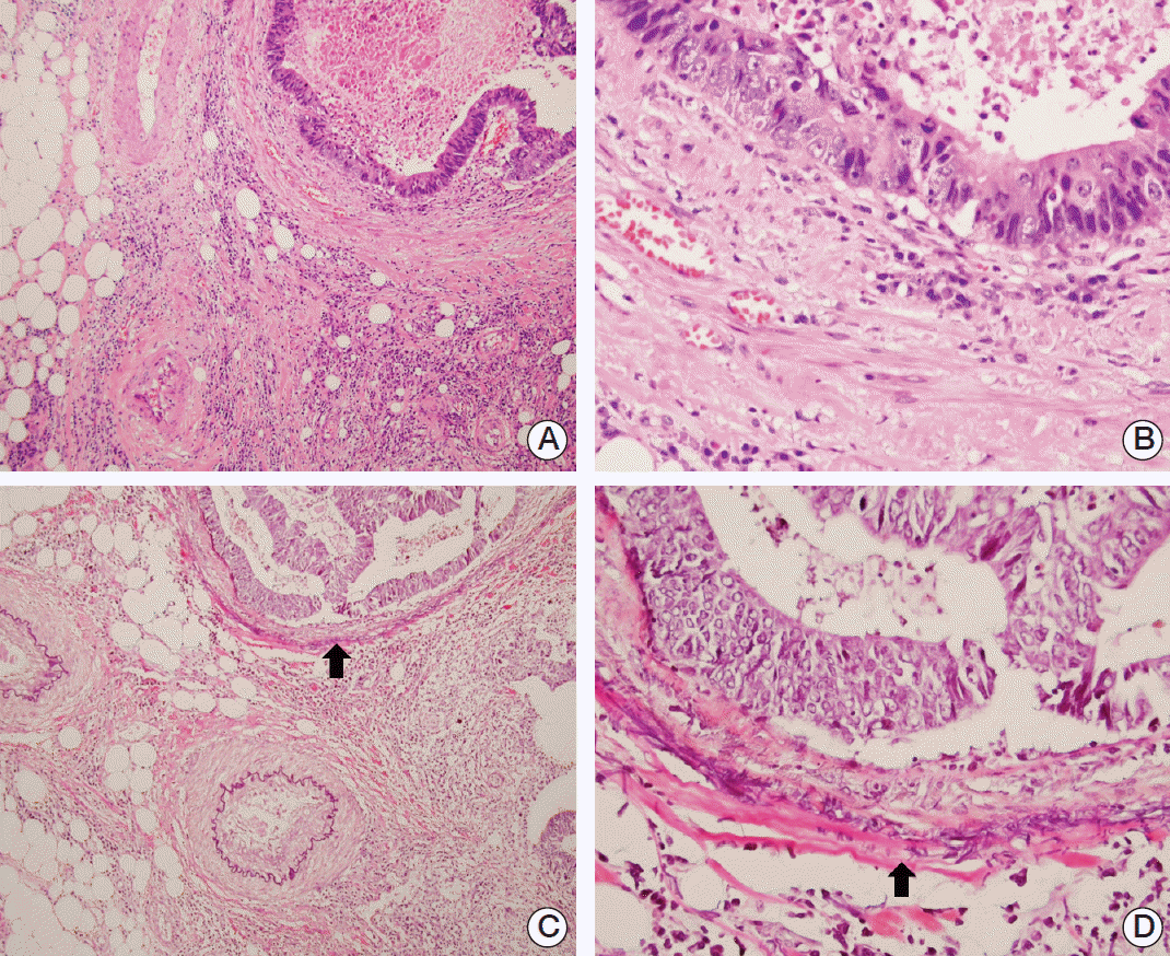
We analyzed extramural VI (VI beyond muscularis propria) with prognostic significance [1,6]. VI was assessed in the 93 included cases of CRC in three steps: (1) evaluation of the original surgical pathology report, (2) re-evaluation of all H&E-stained slides by two pathologists with close attention to morphologic features implying VI as described by Kirsch et al. [14], and (3) re-evaluation of equivocal features based on H&E slides using elastic stains. Regarding morphologic features implying VI, we evaluated the protruding tongue sign (smooth oval-shaped protrusion of tumor cells into periserosal fat tissue near an artery) and the orphan arteriole sign (round tumor nodule near an artery without an accompanying vein) (Fig. 1). For elastic stain, elastic staining was performed using a Roche Elastic Staining Kit (860-005, Ventana Medical Systems Inc., Tucson, AZ) according to the manufacturer’s instructions in all 93 cases of CRCs. One tissue block with equivocal features of VI on H&E stained slides was selected.
The data were analyzed using the Student's t test, Fisher exact test, or the chi-square test for differences between groups. The relationships between different detection methods were assessed using a Spearman rank correlation coefficient. Agreement between different detection methods was assessed with a kappa (κ) value statistics. A value of p < 0.05 was considered statistically significant. Statistical calculations were performed using SSPS ver. 21.0 (IBM Co., Armonk, NY).
Results
Overall, the detection rate of VI was significantly increased as follows: 14/93 in the original pathology report (15.1%), 38/93 in review of H&E slides (40.9%) with attention to the protruding tongue and orphan arteriole signs, and 45/93 (48.4%) using elastic stain, respectively (Table 1). The tumor nodule with a protruding tongue or orphan arteriole sign was identified. When searching under H&E stain, endothelial cells or the smooth muscular layer of a vessel around the tumor nodule were not identified. However, elastic staining showed the elastic layer of a vein around the tumor nodule with the protruding tongue or orphan arteriole signs (Figs. 2 and 3).
Detection of VI on the basis of morphologic features (protruding tongue and orphan arteriole) showed 77.8% sensitivity, 91.1% specificity, 92.1% positive predictive value, 81.8% negative predictive value, a positive likelihood ratio of 12.44 (95% confidence interval [CI], 4.11 to 37.64), and a negative likelihood ratio of 0.24 (95% CI, 0.14 to 0.41). In addition, detection of VI with special attention to morphologic features showed a linear correlation (Spearman correlation coefficient, 0.727; p < 0.001) and with VI detected by elastic stain as the gold standard (Table 1). In comparison, detection of VI on the original pathology report showed only 33.1% sensitivity, 100.0% specificity, 100.0% positive predictive value, 60.8% negative predictive value, and a negative likelihood ratio of 0.69 (95% CI, 0.57 to 0.84). Also, detection of VI with the original pathology report showed a linear correlation (Spearman correlation coefficient, 0.435; p < 0.001) with VI detected by elastic stain as the gold standard (Table 1). In addition, improved agreement was observed between detection methods (detection on the basis of morphologic features, κ=0.719 vs. original pathology report, κ=0.318) using kappa statistics. These data indirectly suggest that morphologic findings (protruding tongue and orphan arteriole signs) are a better method of detection and could enhance the rate of detection of VI in CRC patients. The presence of VI identified by elastic stain was associated with lymph node metastasis, lymphatic emboli, and perineural invasion of tumor cells in T3 or T4 CRCs (Table 2).
In evaluation of the numbers of tissue blocks and number of tissue blocks/tumor diameter (cm) for the primary tumor mass between VI-negative and VI-positive CRCs by elastic stain, there were no differences in the numbers of tissue blocks or number of tissue blocks/tumor diameter (cm) (6.64±0.41 vs. 6.62±0.32, p=0.964; 1.20±0.09 vs. 1.31±0.07, p=0.341).
Discussion
In this study, we demonstrate that the detection rate of VI was significantly increased in review of H&E slides with attention to the “protruding tongue” and “orphan arteriole” signs as well as the use of elastic stain, and we recommend evaluation of these morphological findings for better identification of VI in CRCs in routine surgical practice. According to many reports, VI, or “large vessel” invasion, is an independent poor prognostic factor for CRCs [3-5], and detection of VI in stage II CRC suggests the need for prompt adjuvant chemotherapy by oncologists [6]. Therefore, the VI status should be included in the surgical pathology report of CRC patients by the various CRC reporting protocols, including those of the Royal College of Pathologists (RC Path, London, UK), the Royal College of Pathologists of Australia (RCPA), the CAP, the Japanese Society of Cancer of the Colon and Rectum, and the Korean Standardized Pathology Report for Colorectal Cancer [1,2,16-18].
There are two problems related to VI detection in routine surgical practice. The first is the marked variability in the detection rate of VI, and the other is the lack of consensus guidelines regarding detection methods for VI in CRC. In relation to the variability in the detection rate of VI, there are many contributing factors, including the experience or subspecialty of pathologists, diagnostic criteria, numbers of blocks, and usage of special stains [7-10]. Studies from Ontario, Canada reported these problems and suggested ways to overcome these complicated issues [11,14,15]. Messenger et al. [11] reported that pathologists with a university-affiliated center, a GI pathology subspecialist interest, and acceptance of the “orphan arteriole” sign were associated with a VI detection rate above 10% [11]. In accordance with the above reports, our data showed an improved detection rate of VI with review of H&E slides with specific attention to the “protruding tongue” and “orphan arteriole.”
Kirsch et al. [14] reported that intraobserver agreement between GI and non-GI pathologists was improved with the use of elastic stain in VI detection in CRCs. Messenger et al. [19] and Dawson et al. [20] recommend routine elastic staining on all tumor blocks or on blocks that show the full thickness of the tumor. However, if elastic staining is ordered after slide review, additional costs and efforts are entailed, and turnaround times are delayed. If possible, it is recommended that elastic staining be requested at the time of grossing. Our data showed that detection of VI with morphologic features (protruding tongue and orphan arteriole signs) showed 77.8% sensitivity and 91.1% specificity with elastic stain as the gold standard and improved agreement between detection methods. These data indirectly suggest that use of morphologic findings (protruding tongue and orphan arteriole signs) is a comparable method and could enhance the detection rate of VI in CRC patients. However, 22.2% of cases (10/45 cases) showed false negative results (negative morphologic findings which showed positive on elastic stain), whereas 6.3% of cases (3/48 cases) showed false-positive results (positive morphologic findings which showed negative on elastic stain). It is suggested that more studies regarding morphologic clues are needed to clarify these issues in daily surgical pathology practice of CRC resection specimens.
Conclusion
Taken together, the published body of work and our data recommend that pathologists should perform a careful review of H&E slides in CRC cases with special attention to the protruding tongue and orphan arteriole signs in routine surgical practice and that elastic staining should be considered for equivocal cases. In addition, studies from Ontario, Canada suggest the benefits of national or multi-institutional educational programs (as knowledge transfer on VI detection at a national level) to improve VI detection rates and reduce interobserver variability in Korea.


