





Skip Navigation
Skip to contents

Cancer Res Treat : Cancer Research and Treatment

OPEN ACCESS

Articles

Page Path
HOME > Cancer Res Treat > Volume 47(3); 2015 > Article
Case Report Usefulness of Digital Tomosynthesis for the Detection of Airway Obstruction: A Case Report of Bronchial Carcinosarcoma
Sung-Joon Park, MD1, Ji Yung Choo, MD, PhD1, Ki Yeol Lee, MD, PhD1, Je-Hyeong Kim, MD, PhD2, Jung-Woo Choi, MD, PhD3, Suk Keu Yeom, MD, PhD1, Baek Hyun Kim, MD, PhD1
Cancer Research and Treatment : Official Journal of Korean Cancer Association 2015;47(3):544-548.
DOI: https://doi.org/10.4143/crt.2013.220
Published online: September 11, 2014

1Departments of Radiology, Korea University Ansan Hospital, Korea University of College of Medicine, Ansan, Korea

2Departments of Pulmonology, Korea University Ansan Hospital, Korea University of College of Medicine, Ansan, Korea

3Departments of Pathology, Korea University Ansan Hospital, Korea University of College of Medicine, Ansan, Korea

Correspondence: Ji Yung Choo, MD, PhD Department of Radiology, Korea University Ansan Hospital, Korea University of College of Medicine, 123 Jeokgeum-ro, Danwon-gu, Ansan 425-707, Korea 
Tel: 82-31-412-5228 Fax: 82-31-412-5224 E-mail: quellquell@naver.com
• Received: October 28, 2013   • Accepted: February 11, 2014

Copyright © 2015 by the Korean Cancer Association

This is an Open Access article distributed under the terms of the Creative Commons Attribution Non-Commercial License (http://creativecommons.org/licenses/by-nc/3.0/) which permits unrestricted noncommercial use, distribution, and reproduction in any medium, provided the original work is properly cited.

  • 20,177 Views
  • 68 Download
  • 6 Web of Science
  • 6 Crossref
  • 7 Scopus
prev
Bronchopulmonary carcinosarcoma is a rare malignancy accounting for less than 0.2% to 0.3% of bronchopulmonary tumors. It has admixed components of both malignant epithelial and mesenchymal elements (carcinomatous parenchyma and sarcomatous stroma) [1]. Compared to conventional chest radiography, tomosynthesis is a more sensitive and accurate diagnostic method with a relatively lower radiation dose. Tomosynthesis can provide stereoscopic images and remove the shadows of bones and overlying structures, giving superior visibility of lesions that are difficult to be recognized on chest radiography. We present the first case in which digital tomosynthesis (DTS) was useful for the evaluation of airway obstruction by bronchial carcinosarcoma that was overlooked on initial chest radiography.
A 45-year-old male construction worker for a fire extinguishing facility was admitted to our hospital with a 3-month history of NYHA class II dyspnea. He had previously never been hospitalized and had no underlying disease.
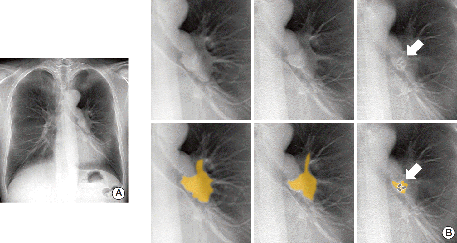
The physician performing the physical examination noticed a wheezing sound on auscultation when the patient breathed. The patient underwent routine chest radiography (Fig. 1). However, we did not detect the airway obstruction on chest radiography at a glance. Suspicious of airway obstruction, we recognized the focally-narrowed left main bronchus. Thus, we recommended DTS for evaluation of the airways. The patient underwent DTS (VolumeRAD, GE Healthcare, Chalfont St. Giles, UK) with flat panel detector radiography (Definium 8000, GE Healthcare) as a follow-up (Fig. 2A).
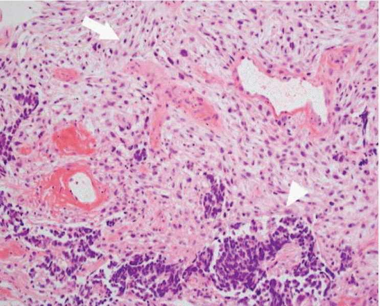
DTS revealed that a lobulating contoured endobronchial lesion nearly obstructed the left main bronchus. Furthermore, we could examine the extent and margins of the endobronchial tumor on anterior-to-posterior coronal images from the DTS (Fig. 2B). After the recognition of the endobronchial mass, the patient underwent computed tomography (CT) scanning for further evaluation. On the CT images, a mildly enhanced finger-like lesion was observed along the left main bronchus and upper lobar bronchus extending to the left lower lobar bronchus (Fig. 3), as suspected on DTS. Subsequent bronchoscopy revealed a mass lesion almost completely occluding the left main bronchus at a site 4 cm below the carina and protruding to the endobronchial space (Fig. 4). A biopsy of the tumor was performed during bronchoscopy and showed that the tumor was composed of epithelial carcinoma that was poorly differentiated, as well as a sarcomatoid component. Positron emission tomography-CT also showed the hypermetabolic status of the lesion and the hilar lymph node, suggesting a malignant process. Surgical procedures were determined based on the evidence presented above. In the surgical field, the main portion of the mass lesion was located in the left main bronchus and left upper lobar bronchus, and a partial portion was located in left lower lobar bronchus. The lesion extended toward the right opposite border of the superior segment of the left lower lobe, so the surgeon performed a left pneumonectomy and mediastinal lymph node dissection. The tumor was 2.5×2 cm and was composed of neuroendocrine carcinoma, adenocarcinoma, squamous cell carcinoma, and sarcoma (Fig. 5). Lymph node metastasis was also reported. The patient did not suffer from any major complications and was put on adjuvant chemotherapy with Genexol and cisplatin.
Bronchopulmonary carcinosarcoma is a rare malignancy that presents as a peripheral solid mass or a central endobronchial lesion. The incidence of the tumor is not fully known due to its rarity, although a few studies have reported that a peripheral type of the carcinosarcoma was found in 62% to 85.7% of total bronchopulmonary carcinosarcomas [2].
Although conventional chest radiography remains in use for the initial imaging of diseases of the chest, it provides poor information for screening the tracheobronchial tree. In the evaluation of tracheobronchial disease, chest radiography is inferior to CT in terms of detection, localization, extent, characterization, and overall information [3]. DTS is a new X-ray application that provides multiple high-resolution slice images of human anatomy including the chest and tracheobronchial tree, while providing more information than radiography and lower radiation doses than CT [4,5]. Airway evaluation by DTS has not yet been reported.
DTS is a refinement of conventional geometric tomography that has been known since the 1930s. DTS acquisition lasts 10 seconds and is comprised of 60 low-dose projections using a tube voltage of 100 kVp, a 1:5 dose ratio, and a 0.3-mm additional Cu filter. The detector is fixed in the standard orthogonal posteroanterior position while the X-ray tube undergoes vertical continuous movement from –17.58° to 17.58°. The 60 projection images obtained are used for the reconstruction of approximately 54 coronal images with a nominal thickness of 4-mm without overlap [5]. The recently reported conversion factor of chest tomosynthesis is 0.26 mSy/Gy/cm2, and dose-area product is 67.45 uGym2 in our case, so the effective dose in our case is 0.18 mSv [6]. The usual effective dose of chest tomosynthesis is 0.12 mSv in the literature, that of chest PA is 0.04 mSv, and that of chest CT is 4 mSv [7]. Thus, the effective dose of DTS is three times more than that of chest X-ray, but 30 times less than that of chest CT scan.
Previous reports on bronchopulmonary carcinosarcoma have investigated its features on imaging or its pathologic findings [8-10]. This case is the first to demonstrate bronchial carcinosarcoma by chest tomosynthesis in the evaluation of an endobronchial lesion that was overlooked on the initial chest radiography in a patient presenting with wheezing. Based on this case, we suggest that endobronchial tumors can be assessed and evaluated by tomosynthesis when there is suspicion of a bronchial problem. Tomosynthesis for the evaluation of airway obstruction can achieve more sensitive detection of endobronchial lesions that may be overlooked on chest radiography, even with low radiation doses compared with CT. This can provide more accurate information than serial follow-up chest radiography for patients with airway obstructive lesions, such as central lung cancer, and can serve as a short-term follow-up modality between relatively long-term CT scans during chemotherapy. The continuous anterior-to-posterior coronal images obtained by DTS can also provide information on the 3-dimensional relationship between the lesion and the bronchial tree, which can give clinicians and surgeons insight into the status of the lesion with low dose radiation.
In conclusion, tomosynthesis can be a modality for the evaluation of airway lesions, providing spatial relationship information with lower radiation doses than ever before.

Conflict of interest relevant to this article was not reported.

Fig. 1.
Initial chest radiography. The endobronchial mass with left upper lobar bronchial narrowing was overlooked on the initial chest radiography.
crt-2013-220f1.gif
Fig. 2.
Digital tomosynthesis (DTS). (A) DTS reveals a lobulating contoured endobronchial lesion nearly obstructing the left main bronchus. (B) The tumor extends to the left upper lobar bronchus and the left lower lobar bronchus. However, the superior segmental bronchus is not obliterated on the last image with patent three holes (arrows). The extent and margins of the endobronchial tumor are well demarcated on serial anterior-to-posterior coronal images from the DTS. The yellow overlay and gray patent bronchi are demonstrated in the lower images.
crt-2013-220f2.gif
Fig. 3.
On the computed tomography images, a mildly enhanced lesion with finger-like appearance was noted along the left main bronchus extending to the upper and lower lobar bronchi, as suspected on digital tomosynthesis.
crt-2013-220f3.gif
Fig. 4.
Bronchoscopic finding. Subsequent bronchoscopy revealed a mass lesion almost completely occluding the left main bronchus at a site 4 cm below the carina and protruding to the endobronchial space. Bronchoscopic biopsy was performed.
crt-2013-220f4.gif
Fig. 5.
Pathologic finding demonstrated sarcomatous (arrow) and carcinomatous (arrowhead) components (H&E staining, ×200).
crt-2013-220f5.gif
  • 1. Huwer H, Kalweit G, Straub U, Feindt P, Volkmer I, Gams E. Pulmonary carcinosarcoma: diagnostic problems and determinants of the prognosis. Eur J Cardiothorac Surg. 1996;10:403–7. ArticlePubMed
  • 2. Sokucu SN, Kocaturk C, Urer N, Sonmezoglu Y, Dalar L, Karasulu L, et al. Evaluation of six patients with pulmonary carcinosarcoma with a literature review. ScientificWorldJournal. 2012;2012:167317PubMedPMC
  • 3. Chen Q, Goo JM, Seo JB, Chung MJ, Lee YJ, Im JG. Evaluation of tracheobronchial diseases: comparison of different imaging techniques. Korean J Radiol. 2000;1:135–41. ArticlePubMedPMC
  • 4. Bath M, Svalkvist A, von Wrangel A, Rismyhr-Olsson H, Cederblad A. Effective dose to patients from chest examinations with tomosynthesis. Radiat Prot Dosimetry. 2010;139:153–8. ArticlePubMed
  • 5. Kim EY, Chung MJ, Lee HY, Koh WJ, Jung HN, Lee KS. Pulmonary mycobacterial disease: diagnostic performance of low-dose digital tomosynthesis as compared with chest radiography. Radiology. 2010;257:269–77. ArticlePubMed
  • 6. Vult von Steyern K, Bjorkman-Burtscher IM, Weber L, Hoglund P, Geijer M. Effective dose from chest tomosynthesis in children. Radiat Prot Dosimetry. 2014;158:290–8. ArticlePubMed
  • 7. Vikgren J, Zachrisson S, Svalkvist A, Johnsson AA, Boijsen M, Flinck A, et al. Comparison of chest tomosynthesis and chest radiography for detection of pulmonary nodules: human observer study of clinical cases. Radiology. 2008;249:1034–41. ArticlePubMed
  • 8. Kim KI, Flint JD, Muller NL. Pulmonary carcinosarcoma: radiologic and pathologic findings in three patients. AJR Am J Roentgenol. 1997;169:691–4. ArticlePubMed
  • 9. Yoshino N, Kubokura H, Yamauchi S, Ohaki Y, Koizumi K, Shimizu K. A true pulmonary carcinosarcoma that required diagnostic differentiation from a pleomorphic adenoma: a case report. Ann Thorac Cardiovasc Surg. 2009;15:42–5. PubMed
  • 10. Zehani A, Ayadi-Kaddour A, Mlika M, Hamrouni R, Fkih L, Marghli A, et al. Primary pulmonary carcinosarcoma. Tunis Med. 2013;91:287–9. PubMed

Figure & Data

REFERENCES

    Citations

    Citations to this article as recorded by  
    • A Rare Case of Tracheal Leiomyoma: Role of Digital Tomosynthesis in Diagnosis and Treatment
      Soo Won Nam, Yeon Joo Jeong, Geewon Lee, Ji Won Lee, Jung Seop Eom, Chang Hun Lee, So Min Park
      Journal of the Korean Society of Radiology.2020; 81(1): 225.     CrossRef
    • Accuracy of Digital Tomosynthesis of the Chest in Detection of Interstitial Lung Disease Comparison With Digital Chest Radiography
      Temphon Kruamak, Rachael Edwards, Stephanie Cheng, Daniel S. Hippe, Ganesh Raghu, Sudhakar N. J. Pipavath
      Journal of Computer Assisted Tomography.2019; 43(1): 109.     CrossRef
    • High‐speed slot‐scanning radiography using small‐angle tomosynthesis: Investigation of spatial resolution
      Christoph Luckner, Magdalena Herbst, Thomas Weber, Marcel Beister, Ludwig Ritschl, Steffen Kappler, Andreas Maier
      Medical Physics.2019; 46(12): 5454.     CrossRef
    • Construction of an Anthropomorphic Phantom for Use in Evaluating Pediatric Airway Digital Tomosynthesis Protocols
      Nima Kasraie, Amie Robinson, Sherwin Chan
      Radiology Research and Practice.2018; 2018: 1.     CrossRef
    • Whole-Body Clinical Applications of Digital Tomosynthesis
      Haruhiko Machida, Toshiyuki Yuhara, Mieko Tamura, Takuya Ishikawa, Etsuko Tate, Eiko Ueno, Katelyn Nye, John M. Sabol
      RadioGraphics.2016; 36(3): 735.     CrossRef
    • A comparison of digital tomosynthesis and chest radiography in evaluating airway lesions using computed tomography as a reference
      Ji Yung Choo, Ki Yeol Lee, Ami Yu, Je-Hyeong Kim, Seung Heon Lee, Jung Won Choi, Eun-Young Kang, Yu Whan Oh
      European Radiology.2016; 26(9): 3147.     CrossRef

    • PubReader PubReader
    • ePub LinkePub Link
    • Cite
      CITE
      export Copy Download
      Close
      Download Citation
      Download a citation file in RIS format that can be imported by all major citation management software, including EndNote, ProCite, RefWorks, and Reference Manager.

      Format:
      • RIS — For EndNote, ProCite, RefWorks, and most other reference management software
      • BibTeX — For JabRef, BibDesk, and other BibTeX-specific software
      Include:
      • Citation for the content below
      Usefulness of Digital Tomosynthesis for the Detection of Airway Obstruction: A Case Report of Bronchial Carcinosarcoma
      Cancer Res Treat. 2015;47(3):544-548.   Published online September 11, 2014
      Close
    • XML DownloadXML Download
    Usefulness of Digital Tomosynthesis for the Detection of Airway Obstruction: A Case Report of Bronchial Carcinosarcoma
    Image Image Image Image Image
    Fig. 1. Initial chest radiography. The endobronchial mass with left upper lobar bronchial narrowing was overlooked on the initial chest radiography.
    Fig. 2. Digital tomosynthesis (DTS). (A) DTS reveals a lobulating contoured endobronchial lesion nearly obstructing the left main bronchus. (B) The tumor extends to the left upper lobar bronchus and the left lower lobar bronchus. However, the superior segmental bronchus is not obliterated on the last image with patent three holes (arrows). The extent and margins of the endobronchial tumor are well demarcated on serial anterior-to-posterior coronal images from the DTS. The yellow overlay and gray patent bronchi are demonstrated in the lower images.
    Fig. 3. On the computed tomography images, a mildly enhanced lesion with finger-like appearance was noted along the left main bronchus extending to the upper and lower lobar bronchi, as suspected on digital tomosynthesis.
    Fig. 4. Bronchoscopic finding. Subsequent bronchoscopy revealed a mass lesion almost completely occluding the left main bronchus at a site 4 cm below the carina and protruding to the endobronchial space. Bronchoscopic biopsy was performed.
    Fig. 5. Pathologic finding demonstrated sarcomatous (arrow) and carcinomatous (arrowhead) components (H&E staining, ×200).
    Usefulness of Digital Tomosynthesis for the Detection of Airway Obstruction: A Case Report of Bronchial Carcinosarcoma

    Cancer Res Treat : Cancer Research and Treatment
    Close layer
    TOP