Clinical Characteristics and Treatment Outcomes of Childhood Acute Promyelocytic Leukemia in Korea: A Nationwide Multicenter Retrospective Study by Korean Pediatric Oncology Study Group
Article information
Abstract
Purpose
Acute promyelocytic leukemia (APL) is a rare disease in children and there are some different characteristics between children and adult. We aimed to evaluate incidence, clinical characteristics and treatment outcomes of pediatric APL in Korea.
Materials and Methods
Seventy-nine pediatric APL patients diagnosed from January 2009 to December 2016 in 16 tertiary medical centers in Korea were reviewed retrospectively.
Results
Of 801 acute myeloid leukemia children, 79 (9.9%) were diagnosed with APL. The median age at diagnosis was 10.6 years (range, 1.3 to 18.0). Male and female ratio was 1:0.93. Thirty patients (38.0%) had white blood cell (WBC) count greater than 10×109/L at diagnosis. All patients received induction therapy consisting of all-trans retinoic acid and chemotherapy. Five patients (6.6%) died during induction chemotherapy and 66 patients (86.8%) achieved complete remission (CR) after induction chemotherapy. The causes of death were three intracranial hemorrhage, one cerebral infarction, and one sepsis. Five patients (7.1%) suffered a relapse during or after maintenance chemotherapy. The estimated 4-year event-free survival and overall survival (OS) rates were 82.1%±4.4%, 89.7%±5.1%, respectively. The 4-year OS was significantly higher in patients with initial WBC < 10×109/L than in those with initial WBC ≥ 10×109/L (p=0.020).
Conclusion
This study showed that the CR rates and survival outcomes in Korean pediatric APL patients were relatively good. The initial WBC count was the most important prognostic factor and most causes of death were related to serious bleeding in the early stage of treatment.
Introduction
Acute promyelocytic leukemia (APL) is a rare and unique subtype of acute myeloid leukemia (AML) characterized by the presence of the recurrent t(15;17) translocation, and accounts for approximately 5%–10% of childhood AML patients [1–5]. APL is classified as AML-M3 (hypergranular promyelocytic leukemia) according to the French-American-British classification system because it presents characteristic cells containing bundles of Auer rods (‘faggots’) in the cytoplasm. However, microgranular variant (M3v) form with irregular nucleus and hypo-granulatioin was also found in about 20% of APL [5]. Pediatric APL patients are known to have a higher frequency of hyperleukocytosis and the M3v subtype at diagnosis compared with adult APL patients [1,3,5,6].
The fusion of promyelocytic leukemia (PML) gene on chromosome 15 with retinoic acid receptor-alpha (RARα) gene on chromosome 17 expresses the PML-RARα protein, which blocks cell differentiation and interferes with apoptosis and tumor suppression. On the presence of the PML-RARα fusion gene in leukemic cells, all-trans retinoic acid (ATRA) induces differentiation of promyeloblasts into neutrophil and arsenic trioxide (ATO) acts both promyeloblast differentiation and apoptosis [1,3]. With the introduction of ATRA treatment, the survival rate of APL showed significant improvement [5]. Several studies have reported that the treatment of pediatric APL based on ATRA and anthracycline chemotherapy results in outcomes similar to those of adult APL [6–10]. ATO has the advantage of being less toxic compared to standard chemotherapy. Therefore, it is increasingly used in childhood APL as first-line treatment as well as for reinduction therapy after relapse [2,3,11].
Hemorrhagic diathesis at diagnosis or after the initiation of cytotoxic chemotherapy is a unique feature of APL and is known to be due to disseminated intravascular coagulation or hyperfibrinolysis triggered by APL blasts. Early deaths from bleeding in APL are reported in 10%–20% [3,5].
Since APL is a rare disease in children, there have not been many studies on pediatric APL. This study was a nationwide, multicenter, retrospective study with the primary aim of investigating the incidence, clinical characteristics and treatment outcomes of childhood APL in Korea.
Materials and Methods
1. Patients
Seventy-nine pediatric patients (up to the age of 18 years) who were newly diagnosed with APL between January 2009 and December 2016 in 16 tertiary medical centers in Korea were enrolled. All patients were newly diagnosed by bone marrow (BM) examination and confirmed to have t(15;17)(q22;q12) by fluorescent in situ hybridization, reverse transcriptase-polymerase chain reaction (RT-PCR) or cytogenetic analysis. The medical records of patients were retrospectively reviewed.
2. Treatment
Induction therapy consisted of oral ATRA (45 mg/m2/day or 25 mg/m2/day), divided into 2 doses, up to hematological remission, and intravenous infusions of anthracycline-based chemotherapy (idarubicin 12 mg/m2 on days 2, 4, 6, and 8 for a total of 4 doses). Patients in morphological complete remission (CR) or partial remission (PR) received 2 to 4 courses of consolidation chemotherapy consisting of anthracycline with or without cytosine arabinoside (Ara-C) according to each institution’s policy. After consolidation, the patients received maintenance chemotherapy for about 2 years, consisting of oral 6-mercaptopurine (50 mg/m2) daily, methotrexate (15 mg/m2) weekly, and ATRA (same dose as in induction) for 15 days every 3 months.
3. Definitions
The relapse-risk group was classified by white blood cell (WBC) count and platelet count at initial presentation according to Sanz’s score [12]: low-risk group (WBC count ≤ 10×109/L, platelets > 40×109/L), intermediate-risk group (WBC count ≤ 10×109/L, platelets ≤ 40×109/L), and high-risk group (WBC count > 10×109/L).
Morphological CR was defined as presence of less than 5% blast cells and promyelocytes in the BM. PR and no response (NR) were defined when BM study showed more than 5% but less than 25% blasts and 25% or more blasts, respectively. Molecular CR and relapse were defined as the disappearance and reappearance of RT-PCR positivity for the PML-RARα fusion transcript.
Diagnosis of retinoic acid (RA) syndrome was made on clinical grounds by the presence of at least three of the following signs without other causes after administration of ATRA: fever, weight gain, respiratory distress, lung infiltrates, pleural or pericardial effusion, hypotension, and renal failure [5].
4. Statistical analyses
Comparison of categorical variables was performed using the chi-square test or Fisher exact test. Overall survival (OS) was defined as the time from diagnosis to death due to any cause or date of last follow-up for surviving patients. Event-free survival (EFS) was defined as the time from diagnosis to relapse, secondary malignancy, death, or date of last follow-up for surviving patients. The EFS and OS curves were constructed using the Kaplan-Meier method and the survival curves between the groups were compared using the log-rank test. p-values < 0.05 were regarded as statistically significant. All analyses were performed by IBM SPSS ver. 26 (IBM Corp., Armonk, NY).
Results
1. Clinical characteristics of the patients
During the 8 years of the study period, a total 801 patients were diagnosed with pediatric AML in 16 tertiary medical centers of Korea. Among them, 79 (9.9%) were diagnosed with APL. The clinical characteristics of pediatric APL patients are summarized in Table 1. The median age at diagnosis was 10.6 years (range, 1.3 to 18.0 years). Male and female ratio was 1:0.93. The median WBC count at diagnosis was 5.1×10⁹/L (range, 0.56 to 182.6), and the median platelet count was 23×109/L (range, 4 to 136). In the recurrence risk group according to the Sanz’s score, 15 patients (19%) were classified as low-risk, 34 (43%) as intermediate-risk, and 30 (38%) as high-risk. Only one patient showed M3v subtype morphologically, and 13 patients had additional cytogenetic rearrangements in addition to t(15;17)(q22;q12). Seventy patients (88.6%) had a tendency to bleed at presentation, of whom 9 (11.4%) had central nervous system (CNS) bleeding.
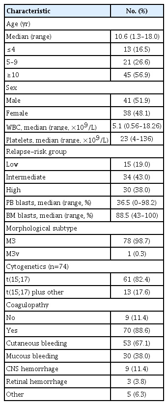
Clinical characteristics of pediatric acute promyelocytic leukemia patients at diagnosis
2. Therapeutic responses
The therapeutic responses are described in Table 2 and Fig. 1. Seventy-six patients received induction treatment, excluding one patient whose follow-up was discontinued after diagnosis and two patients who died of severe bleeding prior to treatment; one was due to intracranial hemorrhage (ICH), and the other was due to pulmonary hemorrhage. Five patients (6.6%) died during induction therapy (3 ICH, 1 cerebral infarction, and 1 sepsis), and 66 (93%) of the remaining 71 patients achieved CR after induction. Of the patients who did not achieve CR, four showed PR and one showed NR. One patient with PR and one patient with NR achieved CR after reinduction with Ara-C, mitoxantrone and oral ATRA. The remaining three patients with PR achieved CR after the first course of consolidation. All 71 patients, including those who showed initial PR and NR, received consolidation treatment, and one patient died from cardiogenic shock during consolidation. All 70 surviving patients continued to show CR status and received maintenance chemotherapy. During and after maintenance chemotherapy, one patient (1.4%) developed secondary malignancy (AML-M2) and five patients (7.1%) experienced relapse. When comparing outcome according to risk group, the CR rate was statistically lower in the low-risk group (low-risk 78.6% vs. intermediate-risk 97% vs. high-risk 95.8%, p=0.019), but the patients with initial PR or NR achieved CR with reinduction or consolidation chemotherapy. There were no differences in recurrence rate, secondary malignancy incidence, and mortality between the 3 groups. The estimated 4-year EFS in pediatric APL was 82.1%±4.4% and the OS was 89.7%±5.1% (Fig. 2). The estimated 4-year OS in the high-risk group was significantly lower than in the other risk groups (79.3%±8.9% vs. 95.9%±3.6%, p=0.020). The 4-year EFS in the high-risk group was slightly lower compared to the other groups, but there was no statistical significance (72.4%±9.1% vs. 87.8%±5.3%, p=0.091) (Fig. 3).
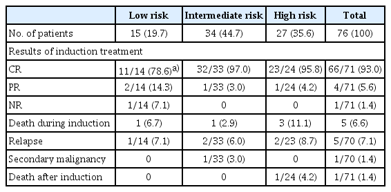
Therapeutic responses according to relapse-risk group in pediatric acute promyelocytic leukemia patients

Flow diagram of patients. APL, acute promyelocytic leukemia; Ara-C, anthracycline with or without cytosine arabinoside; ATRA, all-trans retinoic acid; CR, complete remission; CTx, chemotherapy; F/U, follow-up.
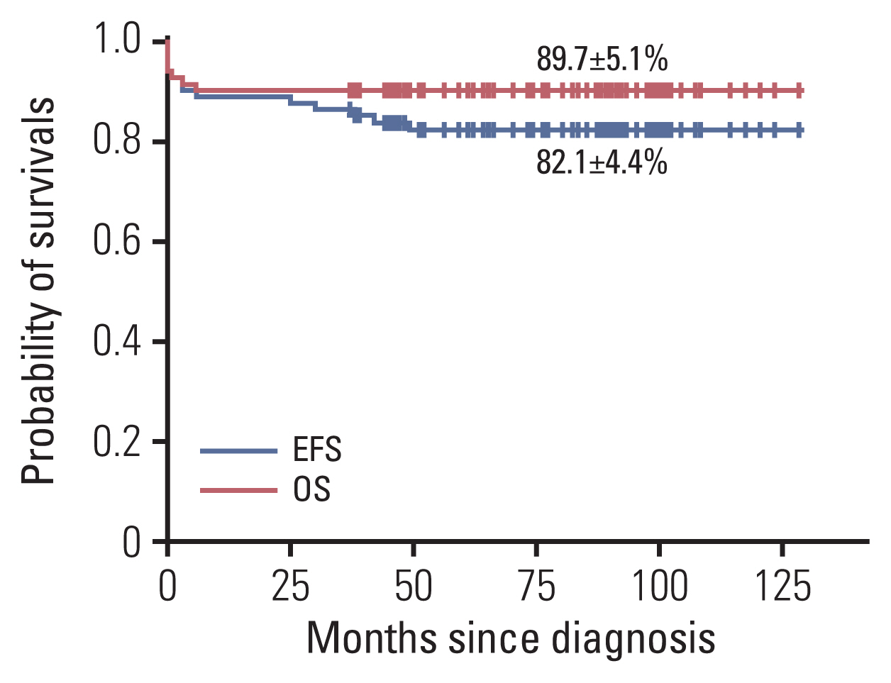
Kaplan-Meier estimates of 4-year overall survival (OS) and event-free survival (EFS) for pediatric acute promyelocytic leukemia.

Kaplan-Meier estimates of 4-year overall survival (A) (p=0.020) and event-free survival (B) (p=0.091) for pediatric acute promyelocytic leukemia according to initial white blood cell (WBC) count.
3. Comparison of treatment responses according to ATRA dosage
Among 76 patients, 51 patients (67.1%) were treated with conventional dose (45 mg/m2/day) ATRA and 25 patients (32.9%) were treated with low-dose (25 mg/m2/day) ATRA. Forty-two patients (55.3%) suffered RA syndrome, and the incidence rate was significantly higher in patients treated with conventional dose compared with those treated with low-dose ATRA (64.7% vs. 36.0%, p=0.018). Twenty-two patients (28.9%) complained of headache after ATRA treatment, and the incidence was higher in patients treated with conventional dose, but there was no statistical significance. No patients were diagnosed with pseudotumor cerebri (PTC). There was no statistical difference between the two groups in CR rate, relapse rate, and survival rate (Table 3, Fig. 4).
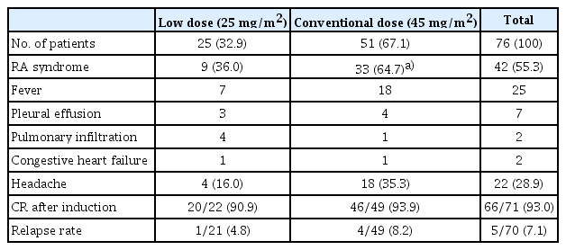
Comparison of ATRA toxicities and responses according to ATRA dosage in pediatric acute promyelocytic leukemia
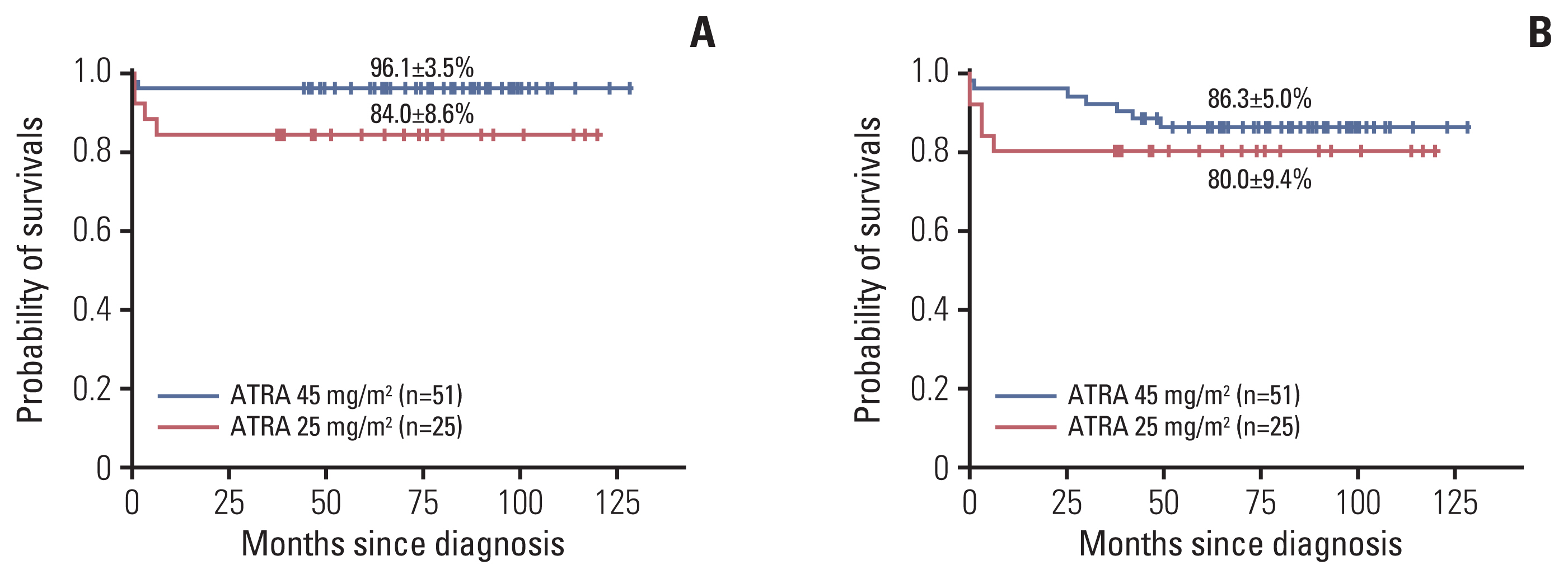
Kaplan-Meier estimates of 4-year overall survival (A) (p=0.069) and event-free survival (B) (p=0.394) for pediatric acute promyelocytic leukemia according to all-trans retinoic acid (ATRA) dosage.
4. Outcomes after relapse
The clinical characteristics and therapeutic outcomes of five relapsed patients are summarized in Table 4. At the time of diagnosis, one patient was in the low-risk group, two in the intermediate-risk group, and two patients were in the high-risk group. The median first CR duration was 38 months (range, 24 to 48 months). All patients achieved second CR after ATO alone (n=3), ATRA combined with ATO (n=1), and ATRA combined with anthracycline (n=1). After achieving second CR, two patients received autologous hematopoietic stem cell transplantation (HSCT), two patients received allogeneic HSCT, and one patient is scheduled for allogeneic HSCT. One patient relapsed again in the BM and CNS 12 months after autologous HSCT, and after achieving a third CR by previously used ATRA alone, he received allogeneic HSCT from a human leukocyte antigen matched related sibling donor. All of the relapsed patients are alive without evidence of subsequent disease relapse.
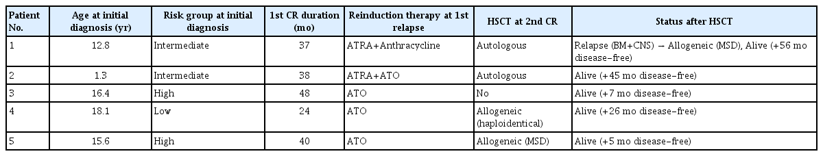
Treatment outcome after relapse in five patients
Discussion
The present retrospective, nationwide, multicenter study evaluated the incidence and survival outcomes of APL in Korean children. The incidence of pediatric APL in Korea was about 10% of de novo AML patients less than 18 years old in this study. The frequency of APL was similar to results of other studies that have been reported [6–9]. The age distribution of patients with APL different from that of other AML patients. While APL is uncommon in the first decade of life, the incidence increases during the second decade reaching a plateau during early adulthood, and diagnosis at age < 1 year is very rare [3,10]. The sex ratio of childhood APL varies with study size, and as the study size increased, sex differences became less apparent [4]. In our study, the median age at diagnosis was 10.6 years (range, 1.3 to 18.0 years), and the male to female ratio was almost the same.
Initial leukocyte count is known as the most important factor in APL, and the patients with an initial WBC count of ≥ 10×109/L are classified as a high-risk group for recurrence [9,12,13]. Compared with adult APL patients, pediatric APL patients more frequently present hyperleukocytosis at diagnosis (approximately 40% in children vs. 20%–25% in adults) and are more likely to have the M3v subtype [3,10]. Nevertheless, several studies of pediatric APL have reported similar survival rates to those of adult APL [6–9]. In our study, 38% of patients were classified as high risk, but M3v was observed in only one patient.
Since simultaneous administration of ATRA and anthracycline-containing chemotherapy was established as frontline treatment for APL, favorable outcomes have been reported in several multicenter studies of pediatric APL [6–9]. The GIMEMA-AIEOP group studied 124 pediatric APL patients and reported a CR rate of 96%, and the OS and the EFS of 89% and 76%, respectively [9]. In the PETHEMA group, 66 children were included and reported a CR rate of 92%, and OS and the disease-free survival (DFS) rates of 87% and 82%, respectively [7]. In a recent ICC-APL-01 trial, the OS and the EFS of 258 pediatric patients were reported as 94.6% and 79.9%, respectively [14]. In our study, the survival rates of patients treated with a combination of ATRA and anthracycline-containing chemotherapy were similar to other previously reported results (OS, 89.7±5.1%; EFS, 82.1±4.4%). Although the CR rate of the low-risk group in this study was less than was found in the other risk groups, the final OS and EFS rates were high as the patients who initially achieved PR or NR patients in this group finally achieved CR with reinduction or consolidation chemotherapy.
In the previous study by the PETHEMA group, the patients in the high-risk group had a higher cumulative incidence of relapse (CIR) and a lower DFS than the other groups (CIR 31% vs. 3.5%, p=0.01; DFS 68±24% vs. 96±7%, p=0.01) [7]. Another study by GIMEMA-AIEOP group also reported that the EFS in the high-risk group was significantly lower than in the other groups (59% vs. 83%, p=0.007) [9]. In this study, the EFS and the OS in the high-risk group was worse than in the other groups, and the initial high leukocyte count was the only prognostic factor (EFS, 72.4±9.1% vs. 87.8±5.3%, p=0.091; OS, 79.3±8.9% vs. 95.9±3.6%, p=0.020).
One of the major complications of APL is a life-threatening consumptive coagulopathy. This coagulopathy usually develops early in the course of disease before starting APL treatment or within a few weeks, and the CNS and the lungs are the most common areas of fatal bleeding [2,5,15,16]. In the COG AAML0631 trial, they evaluated 79 pediatric patients, and four early deaths (all high-risk group) and 13 non-fatal but clinically significant coagulopathy events were observed before the end of induction [15]. Of the 258 patients enrolled in the ICC-APL-01 trial, eight early deaths (1 standard risk and 7 high risk) were observed, and the cause of death was ICH in all eight patients [14]. In our study, seven (8.3%) early deaths were observed; six patients died from bleeding due to coagulopathy, and one patient died from sepsis. Although one patient died due to cardiogenic shock after the end of induction, almost all deaths occurred prior to treatment or before the end of induction. In addition, four out of six early deaths due to coagulopathy were in the high-risk group, which was a major contributing factor in the significantly reduced OS rate in the high-risk group in this study. Early recognition of disease and early treatment with ATRA will decrease early deaths from disease in APL children.
The side effects of ATRA treatment include RA syndrome, headache, and PTC. RA syndrome is known to be another leading cause of early death in APL, and several studies have shown that the incidence of RA syndrome in pediatric APL is about 10%–20%, similar to that of adult APL [2,6–9]. On the other hand, other side effects related to ATRA, such as headache and PTC, have been reported to occur more frequently in children than in adults [6,9]. In order to reduce these complications, studies on the use of low-dose ATRA were conducted, and one study reported that there was no difference between 25 and 45 mg/m2/day of ATRA in the therapeutic efficacy and pharmacokinetic results [17]. For this reason, many protocols for the treatment of pediatric APL have since used ATRA at a dose of 25 mg/m2/day, and the treatment outcomes were reported to be similar to those who received conventional dose ATRA [7,9,14]. In our study, the incidence of RA syndrome and headache was 55.3% and 28.9%, respectively, which was higher than reported in other studies. Specifically, the incidence of ATRA side effects was significantly higher in patients treated with 45 mg/m2/day of ATRA than those treated with 25 mg/m2/day of ATRA. But almost all cases of side effects including RA syndrome were transient, reversible, non-fatal and there was no significant difference in the OS and the EFS between groups according to ATRA dosage.
More recently, several studies reported that ATO therapy was effective and well-tolerated in pediatric APL [8,11,18,19]. Moreover, ATO could allow reduction of the overall amount of conventional chemotherapy and the cumulative dose of anthracyclines. In studies of relapsed pediatric APL patients, ATRA+chemotherapy or ATO alone have been reported to be effective in obtaining CR as a salvage therapy [8,18,20]. Especially, ATO is well documented to induce second remission, in up to 80% of patients after two courses. Therefore, ATO containing regimens are the preferred salvage therapy [11]. Five patients who relapsed in our study also successfully obtained CR with salvage treatment. Four patients received ATO with or without ATRA for induction therapy; it was tolerable and effective in obtaining second CR in relapsed patients. In addition, one patient who experienced second relapse after autologous HSCT achieved third CR after induction therapy with previously used ATRA alone.
Regardless of the agents used to induce second CR, HSCT is necessary to prevent further recurrence in relapsed APL children. Dvorak et al. [21] analyzed the results of 32 pati-ents who received HSCT (11 autologous, 21 allogeneic) for relapsed or refractory APL. There was no statistical difference in survival rate between autologous and allogeneic HSCT. But, autologous HSCT was associated with a lower incidence of treatment-related mortality, while allogeneic HSCT was associated with a lower relapse rate. In our study, 1 (patient No. 1) out of two patients who received autologous HSCT experienced relapse again and eventually received an allogeneic HSCT from an human leukocyte antigen (HLA) matched sibling. All of the five relapsed patients are alive without evidence of disease. Like other types of leukemia, allogeneic HSCT may enhance survival because of the graft-versus-leukemia effect.
In conclusion, our study showed that the CR rate and OS in Korean pediatric APL patients were comparable to results that have been previously reported by study groups from North America and Europe. The initial WBC count was the most important prognostic factor in APL, and most of the causes of death were serious bleeding complications in the early stage of treatment. Low-dose ATRA was effective in reducing incidence of RA syndrome while resulting in survival outcomes comparable to conventional dose ATRA. In order to define other significant risk factors of APL in children, large scaled randomized prospective trials will be needed.
Notes
Ethical Statement
This study was approved by the institutional review board (IRB) of the Pusan National University Yangsan Hospital, Korea (IRB No. 04-2019-021). Written informed consents were waived because of the retrospective nature of this study.
Author Contributions
Conceived and designed the analysis: Park KM, Yoo KH, Kim SK, Lee JW, Chung NG, Ju HY, Koo HH, Lyu CJ, Han SM, Han JW, Choi JY, Hong KT, Kang HJ, Shin HY, Im HJ, Koh KN, Kim H, Kook H, Baek HJ, Kim BR, Yang EJ, Lim JY, Park ES, Choi EJ, Park SK, Lee JM, Shin YJ, Kim JY, Park JK, Kong SK, Choi YB, Lim YT, Cho B.
Collected the data: Park KM, Yoo KH, Kim SK, Lee JW, Chung NG, Ju HY, Koo HH, Lyu CJ, Han SM, Han JW, Choi JY, Hong KT, Kang HJ, Shin HY, Im HJ, Koh KN, Kim H, Kook H, Baek HJ, Kim BR, Yang EJ, Lim JY, Park ES, Choi EJ, Park SK, Lee JM, Shin YJ, Kim JY, Park JK, Kong SK, Choi YB, Lim YT, Cho B.
Contributed data or analysis tools: Park KM, Yoo KH, Kim SK, Lee JW, Chung NG, Ju HY, Koo HH, Lyu CJ, Han SM, Han JW, Choi JY, Hong KT, Kang HJ, Shin HY, Im HJ, Koh KN, Kim H, Kook H, Baek HJ, Kim BR, Yang EJ, Lim JY, Park ES, Choi EJ, Park SK, Lee JM, Shin YJ, Kim JY, Park JK, Kong SK, Choi YB, Lim YT, Cho B.
Performed the analysis: Park KM, Yoo KH, Kim SK, Lee JW, Chung NG, Ju HY, Koo HH, Lyu CJ, Han SM, Han JW, Choi JY, Hong KT, Kang HJ, Shin HY, Im HJ, Koh KN, Kim H, Kook H, Baek HJ, Kim BR, Yang EJ, Lim JY, Park ES, Choi EJ, Park SK, Lee JM, Shin YJ, Kim JY, Park JK, Kong SK, Choi YB, Lim YT, Cho B.
Wrote the paper: Park KM, Yoo KH, Kook H, Lim YT, Cho B.
Conflicts of Interest
Conflict of interest relevant to this article was not reported.
Acknowledgments
This study was supported by funding from the Korea Childhood Leukemia Foundation in 2019.