Nationwide Trends in the Incidence of Melanoma and Non-melanoma Skin Cancers from 1999 to 2014 in South Korea
Article information
Abstract
Purpose
This descriptive study was aimed to examine trends in the incidence of melanoma and nonmelanoma in South Korea.
Materials and Methods
The nationwide incidence data for melanoma and non-melanoma skin cancer was obtained from the Korea Central Cancer Registry. Age-standardized rates were calculated and analyzed, using a Joinpoint regression model.
Results
The incidence of basal cell carcinoma has increased dramatically both in men (average annual percentage change [AAPC], 8.0 [95% confidence interval (CI), 6.0 to 10.1]) and women (AAPC, 9.0 [95% CI, 7.5 to 10.4]). Squamous cell carcinoma has also steadily increased both in men (AAPC, 3.3 [95% CI, 2.6 to 4.0]) and women (AAPC, 6.8 [95% CI, 5.3 to 8.4]). Cutaneous melanoma increased continuously from 1999 to 2014 inwomen (AAPC, 3.5 [95% CI, 2.4 to 4.6]), whilst rapidly increasing in men until 2005 (APC, 7.9 [95% CI, 2.4 to 13.7]) after which no increase has been observed (APC, ‒0.2 [95% CI, ‒2.3 to 2.0]).
Conclusion
The incidence rates of melanoma and non-melanoma skin cancer have increased over the past years, with the exception of melanoma in men. Further studies are required to investigate the reasons for the increased incidence of these skin cancers in South Korea.
Introduction
Skin cancer including melanoma and non-melanoma skin cancer is one of the most commonly diagnosed types of cancer worldwide. In the United States, cutaneous melanoma is expected to be the fifth leading cancer type in men and the sixth in women in 2017 [1]. Although the incidence rate of melanoma is much lower than that of basal cell carcinoma or squamous cell carcinoma, melanoma has a much worse prognosis [2].
Although melanoma and non-melanoma skin cancers are commonly diagnosed in Western countries including the United States [2-4], they are rare in Asian countries including Korea [3-6]. In addition, incidence rates of melanoma in the United States vary with respect to race and ethnicity [7]. Although studies have observed an increase in the incidence of melanoma and non-melanoma skin cancer in Western countries [3,4,7-11], some have reported that the incidence among Asians and Pacific Islanders remain stable [4,7].
Few studies on the trends in the incidence of melanoma or non-melanoma skin cancer among East Asian, which was known as having low incidence rate compared to that of Caucasian [3-6]. In addition, differences have been observed in the incidence of non-melanoma skin cancer even within the same Asian population [6,12,13].
The Korea Central Cancer Registry (KCCR), which covers the entire Korean population, has provided reliable nationwide cancer incidence data since the year 1999 [14]. Although few population-based cancer registries have provided incidence rates of non-melanoma skin cancer, the KCCR has especially collected a nationwide incidence data of nonmelanoma skin cancer including squamous cell carcinoma and basal cell carcinoma as well as cutaneous melanoma. Therefore, this study aimed to examine the trends in incidence of melanoma and non-melanoma skin cancer in Korea, using data from the Korean nationwide cancer registry from 1999 to 2014.
Materials and Methods
1. Data sources
This descriptive study was based on the national cancer registry database of South Korea. The Information about patients diagnosed with skin cancer between 1999 and 2014 was extracted from the Korean national cancer incidence database of the KCCR. The KCCR covered the nationwide cancer incidence in South Korea from 1999 [15]. The completeness of data of the KCCR for 2013 was estimated to be 97.8% [14]. The proportion of cases identified by death certificate only was about 1.1%. The proportions of microscopic verification and unknown age for all skin cancers were 99.3% and 0.0%, respectively, in 2014. Most cancer registries in other countries do not collect information about nonmelanoma cancer. However, the KCCR has consistently provided nationwide incidence data for non-melanoma skin cancer since 1999. Detailed information on the Korean national cancer incidence database has been described elsewhere [14,15].
2. Case definitions
Skin cancer was defined according to site as “C43” and “C44” according to the International Classification of Diseases, 10th revision (ICD-10) code [16]. Melanoma was defined as having a morphology code of “8720-8790.” Skin cancer with a morphology code of “8050-8078” or “8083-8084” was classified as squamous cell carcinoma and skin cancer coded as “8090-8098” was classified as basal cell carcinoma according to the International Classification of Diseases for Oncology, third edition (ICD-O-3) code [17,18]. Skin cancer sites were categorized into sun-exposed sites (face and neck: ICD-10 codes for C430-C433, C440-C443), and sites that are usually not exposed to sun (trunk and lower limbs: ICD-10 codes for C435, C437, C445, C447), to perform subgroup analysis.
3. Statistical analysis
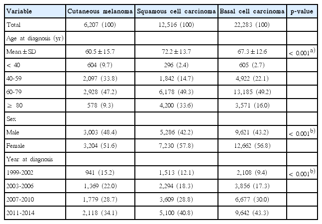
The baseline characteristics of patients with skin cancer are presented according to the histological subgroup in Table 1. The mean age of patients was compared using ANOVA. The number (%) of study participants are described according to the age at diagnosis (< 40, 40-59, 60-79, and ≥ 80 years), sex, and year of diagnosis (1999-2002, 2003-2006, 2007-2010, and 2011-2014). A chi-square test was performed to assess the relationship between categorical variables and histological subgroup of skin cancer.

Baseline characteristics of patients with cutaneous melanoma, squamous cell carcinoma, and basal cell carcinoma in Korea, from 1999 to 2014
Age-standardized incidence rates were calculated as the sum of weighted incidence rate for each 5-year age group using the Segi’s world standard population [19]. The age-standardized incidence rates for melanoma, squamous cell carcinoma, and basal cell carcinoma are expressed as number of new cases per 100,000 people. The Joinpoint regression model is a piecewise linear regression analysis that examines trends in rates and identifies statistically significant changes over time [20]. This model was used to detect the best-fitting points where there are significant changes in the incidence trend for melanoma, squamous cell carcinoma and basal cell carcinoma. Annual percent changes (APC) were used to summarize trends in incidence for each interval. In addition, the average annual percent change (AAPC) was also calculated as a weighted average of APCs to summarize the trend over the entire period between 1999 and 2014. APC and AAPC present percent change and average percent change per 1 year, respectively. Subgroup analysis was performed according to the age group, and the legions were divided into sites exposed to sun and sites that are not.
p-values less than 0.05 were considered statistically significant. All statistical analyses were performed using Stata ver. 12.0 (StataCorp LP, College Station, TX) and SAS ver. 9.3 (SAS Institute Inc., Cary, NC) and Joinpoint regression program ver. 4.1.1 (Surveillance Research Program, National Cancer Institute, Bethesda, MD).
4. Ethical statement
The study was approved by the Institutional Review Board of National Cancer Center (NCC2017-0043) and performed in accordance with the principles of the Declaration of Helsinki. The informed consent was waived.
Results
1. Baseline characteristics
In South Korea, 6,207 cases were diagnosed as cutaneous melanoma, 12,516 cases as squamous cell carcinoma and 22,283 cases as basal cell carcinoma, between 1999 and 2014 (Table 1). The mean age of patients with squamous cell carcinoma and basal cell carcinoma were 72.2±13.7 years and 67.3±12.6 years, respectively. Patients with cutaneous melanoma (60.5±15.7 years) were younger than those with non-melanoma skin cancer (p < 0.001). About 50% of all patients with skin cancer were between the ages of 60 and 79 years. With respect to sex, there were more female patients than male patients, and the proportion of female patients with squamous cell carcinoma (57.8%) and basal cell carcinoma (56.8%) was comparatively higher than that of female patients with cutaneous melanoma (51.6%) (p < 0.001). The number of patients diagnosed with melanoma and nonmelanoma skin cancer has increased over time.
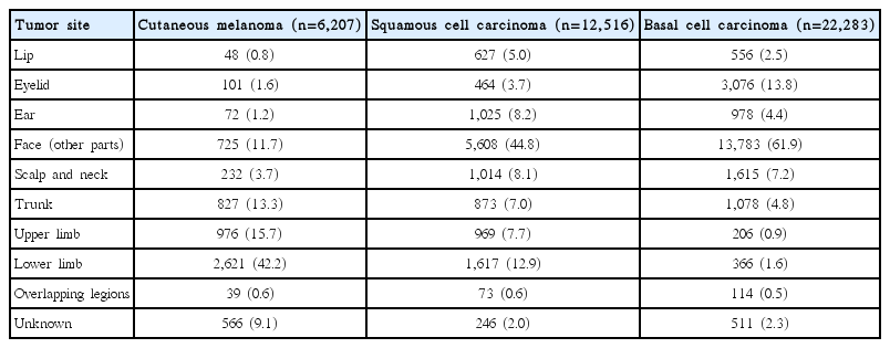
Table 2 shows the distribution of primary tumor sites of cutaneous melanoma, squamous cell carcinoma, and basal cell carcinoma. More than 50% of the non-melanoma skin cancer cases developed on the head and neck including the face, while cutaneous melanoma were mainly detected at the lower limbs (42.2%), followed by the upper limbs (15.7%) and the trunk (13.3%).
2. Incidence rate of melanoma and non-melanoma skin cancer
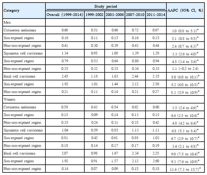
The age-standardized incidence rates of cutaneous melanoma, squamous cell carcinoma, and basal cell carcinoma during 1999-2014 were 0.66, 1.34, and 2.45 per 100,000 people for men, and 0.58, 1.04, and 2.07 per 100,000 for women, respectively (Table 3). During 1999-2002 and 2011-2014, the basal cell carcinoma incidence rate has notably increased from 1.18 to 2.35 in men (AAPC, 8.0 [95% confidence interval (CI), 6.0 to 10.1]) and from 0.98 to 2.25 in women (AAPC, 9.0 [95% CI, 7.5 to 10.4]). The incidence rate of squamous cell carcinoma has increased more rapidly in women (AAPC, 6.8 [95% CI, 5.3 to 8.4]) than in men (AAPC, 3.3 [95% CI, 2.6 to 4.0]). The age-standardized incidence rate of cutaneous melanoma has also increased from 0.51 in 1999-2002 to 0.67 in 2011-2014 among men (AAPC, 3.0 [95% CI, 0.8 to 5.3]), and from 0.43 in 1999-2002 to 0.60 in 2011-2014 among women (AAPC, 3.5 [95% CI, 2.4 to 4.6]).
3. Joinpoint regression analysis for trends in incidence of melanoma and non-melanoma skin cancer
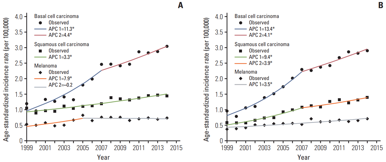
In the results of the Joinpoint regression analysis, the most notable change was observed for basal cell carcinoma from 1999 to 2014 both in men and women (Fig. 1). Basal cell carcinoma has increased dramatically from 1999 to 2007 both in men (APC, 11.3 [95% CI, 7.6 to 15.1]) and women (APC, 13.4 [95% CI, 10.7 to 16.1]). Since 2007, the increasing trend in the age-standardized incidence rate of basal cell carcinoma has been attenuated both in men (APC, 4.4 [95% CI, 1.9 to 7.0]) and women (APC, 4.1 [95% CI, 2.3 to 6.0]). The incidence rate of squamous cell carcinoma has also steadily increased from 1999 to 2014 in men with a 3.3% APC (95% CI, 2.6 to 4.0). In women, the incidence rate of squamous cell carcinoma rapidly increased from 1999 to 2007 (APC, 9.4 [95% CI, 6.7 to 12.3]) until the increasing trend was attenuated from 2007 to 2014 (APC, 3.9 [95% CI, 1.9 to 6.0]). The incidence rate of cutaneous melanoma has increased continuously from 1999 to 2014 in women (APC, 3.5 [95% CI, 2.4 to 4.6]), whereas in men, the increase was rapid until 2005 (APC, 7.9 [95% CI, 2.4 to 13.7]) after which the increase was attenuated (APC, ‒0.2 [95% CI, ‒2.3 to 2.0]).

(A) Trends in age-standardized incidence rates for melanoma, basal cell carcinoma, and squamous cell carcinoma in Korean men, 1999-2014. (B) Trends in age-standardized incidence rates for melanoma, basal cell carcinoma and squamous cell carcinoma in Korean women, 1999-2014. APC, anuual percent change. The age-standardized incidence rates are presented as incidence cases per 100,000 people using Segi’s world standard population as standard population. Joinpoint regression analysis was used to determine whether there were significant changes in trends in age-standardized incidence rates for the period between 1999 and 2014. *p < 0.05.
4. Trends in the incidence rate of melanoma and nonmelanoma by subsites
Subgroup analyses were conducted by categorizing sites into sun-exposed sites and sites that were not exposed to sun. The incidence rate of melanoma consistently increased mainly in the sites that were not exposed to sun (Tables 3 and 4). Although the incidence rate of melanoma has increased in the early period (1999-2002), it remained stable from 2003 to 2014 for both men and women (Table 3). Squamous cell carcinoma incidence rate for the sites exposed to sun had a larger increase than the incidence rate for sites that were not exposed to sun, while basal cell carcinoma has significantly increased regardless of the sun exposure. However, the absolute increase in the incidence rate between the period of 1999-2002 and 2011-2014 was greater in the sites exposed to sun than in sites that were not exposed to sun.
5. Trends in the incidence rate of melanoma and nonmelanoma by age group
Age-stratified analysis was performed to explore whether there were different trends in melanoma and non-melanomas by age group. Of all age groups and sexes, the incidence rates of melanoma, squamous cell carcinoma, and basal cell carcinoma among patients aged ≥ 80 years old showed the largest increase (S1 and S2 Tables), while the incidence rates of melanoma and squamous cell carcinoma among patients aged less than 40 years old did not increase significantly. Interestingly, only basal cell carcinoma showed a significant increasing trend among these patients.
S3 and S4 Tables showed trend of melanoma, basal cell carcinoma and squamous cell carcinoma of face, because face accounted for 61.9% of basal cell carcinoma.
Discussion
Our findings demonstrated that the incidence of cutaneous melanoma, squamous cell carcinoma, and basal cell carcinoma has increased from 1999 to 2014 (except for melanoma skin cancer in men, which was observed to have attenuated during 2005-2014) although these rates happen to be much lower than previously reported among white skinned population.
Although little information on the trends in skin cancer in Asian countries is available, there are several reports about changes in the incidence of melanoma and non-melanoma in Singapore [5,12], Hong-Kong [21], and Japan [22]. However, the incidence of melanoma and non-melanoma shows different trends, depending on the race and the country, even within the population of Asians [12,13].
The incidence of melanoma has increased in most western countries [23]. In the Unites States, the incidence increased from 1992 to 2006 among the white-skinned population for all age groups as well as with respect to tumor thickness [24]. In Denmark, the incidence of invasive melanoma increased by 4.5% in men and by 4.3% in women, from 1985 to 2012 [25]. Contrary to the increasing trends in incidence of melanoma in Western countries, the incidence of melanoma has showed either stable or decreasing trends in Asian countries. The International Agency for Research on Cancer (IARC) reported in a study that the incidence of melanoma in most Asian countries were relatively low and stayed stable [4]. The incidence rate of melanoma also remained stable from 1967 to 1997 in Singapore [5]. Furthermore, there is evidence showing a decline in the incidence rate of melanoma from 1983 through to 2002 in Hong Kong [21].
In our study, sex-related differences in the incidence rate of melanoma were observed. There is evidence that since 2005, the incidence rate of melanoma has been stabilized in men, whereas there has been a slight but steady increase in the incidence of melanoma in women. Although we are uncertain about the exact reason why these trends vary in relation to sex, a previous study has also shown a rapid increase in ocular melanoma in women than in men [26]. Differences in these trends in relation to sex may be partly due to the differences in the medical behavior of men and women. Men have lower participant rates for cancer screening than women [27]. In addition, a study reported that men complained less of symptoms before being diagnosed with uveal melanoma [28]. In our study, we observed that the upper and lower extremities were predominant sites for melanoma. A study demonstrated that cutaneous melanoma occurred mainly at acral sites (such as the palm or sole or nails or toenails) in South Korea [29]. Although these regions are less likely to be exposed to direct sunlight, the effects of increased ultraviolet exposure from other practices such as indoor tanning, could be attributed to the increase in melanoma. Indeed, Surveillance, Epidemiology, and End Results data in the United States showed that incidence rate of melanoma has increased regardless of socioeconomic status. This study suggests that the incidence rate of melanoma may be affected by environmental exposure, such as sunlight or tanning. However, very few people use indoor tanning in South Korea compared to people in western countries. Therefore, the increase in melanoma in South Korea may be mainly attributed to the increase in awareness of skin cancer among general population and dissemination of the punch biopsy tool in South Korea, although absolute increase of cutaneous melanoma was small. Indeed, a population-based study in Germany (SCREEN project) showed that skin cancer screening could increase the incidence of melanoma.
Although there are a few cancer registries that record the nationwide incidence of non-melanoma skin cancer, several studies have reported consistently increasing trends in the incidence of non-melanoma skin cancer. In a systematic review of the worldwide incidence of non-melanoma skin cancer, the incidence rate of basal cell carcinoma, and squamous cell carcinoma showed increasing trends in European men [3]. In Canada, basal cell carcinoma and squamous cell carcinoma increased by 2.4% each year among adults aged ≥ 40 years from the 1970s to 2000 [10]. In the United Kingdom, basal cell carcinoma increased by 39% from 2000 to 2011 in all age groups ≥ 30 years [30].
Although the incidence of both basal cell carcinoma and squamous cell carcinoma has increased in the Western countries, incidence trends of non-melanoma skin cancer varied according to subtype in the Asian countries. A multi-center study in Japan reported that the age adjusted incidence rate of basal cell carcinoma increased from the period 1976-1980 to 1986-1990, but the rate of squamous cell carcinoma did not increase significantly [21]. In Singapore, the incidence rate of basal cell carcinoma has increased by 3.2% per year for men and by 2.8% per year for women. However, the incidence rate of squamous cell carcinoma has decreased by 0.9% per year in both sexes [5]. In accordance with other studies, our findings also showed basal cell carcinoma to have the highest incidence, which increased most rapidly from 1999 to 2014 in both men and women in South Korea. A rapid increase in the incidence of squamous cell carcinoma was also observed in both sexes in South Korea, as reported in Western countries. However, the increasing trends of basal cell carcinoma and squamous cell carcinoma incidence have attenuated since year 2007. Although the most common site of nonmelanoma was the face, which is likely to be exposed to sunlight, it is uncertain whether the increase in non-melanoma skin cancer was due to an increase in the detection of changes in environmental factors such as ultraviolet exposures. In this study, it was difficult to account for the inconsistencies in the trends of non-melanoma skin cancer. Further studies are needed to investigate the reasons for the increase in melanoma and non-melanoma skin cancer in South Korea.
In our study, old age group showed large increasing trends in the incidence rates of melanoma and non-melanoma, while there were different trends in the incidence rates for young age group between squamous cell carcinoma and basal cell carcinoma. For squamous cell carcinoma, there was no significant increase among patients < 40 years old. However, basal cell carcinoma showed significant increasing trends among patients < 40 years old. In the United Kingdom, there was a report that the incidence of basal cell carcinoma has increased between 1981-1989 and 1998-2006 among young people < 30 years old. In Minnesota, incidence rate of basal cell carcinoma among patients < 40 years old has also increased between 1976 and 2003. However, incidence rate of squamous cell carcinoma among patients < 40 years old in Minnesota has increased from 1976 to 1999, but it has leveled off since 1999.
Although our study is based on a representative national cancer registry, it has some limitations that should be considered when interpreting results. Firstly, the increase in the incidence of cancer may be partly due to an improvement in the completeness in the cancer registry. However, the population coverage of the KCCR has been almost 100% since the year 1999. Therefore, the effects of the improvement in completeness of the cancer registry would be probably very small, even if there was an increase in completeness in the KCCR. Secondly, the KCCR did not collect information on tumor characteristics such as thickness of tumor, Clark’s level of invasion, or routes of tumor detection. Therefore, we could not investigate the incidence rate of skin cancer based on the thickness of tumor or routes of tumor detection.
In summary, our study provides reliable nationwide incidence rates of melanoma, squamous cell carcinoma, and basal cell carcinoma in South Korea. Our findings also show an increase in the incidence of squamous cell carcinoma and basal cell carcinoma in both sexes in South Korea, but an increasing trend in the incidence of melanoma was observed only in women. Further studies are needed to investigate the causes of the increased incidence of these skin cancers in South Korea.
Electronic Supplementary Material
Supplementary materials are available at Cancer Research and Treatment website (http://www.e-crt.org).
Notes
Conflict of interest relevant to this article was not reported.
Acknowledgements
This work was supported by the National Cancer Center Grant (Grant number NCC-1610170-2).


