





Skip Navigation
Skip to contents

Cancer Res Treat : Cancer Research and Treatment

OPEN ACCESS

Articles

Page Path
HOME > Cancer Res Treat > Volume 44(3); 2012 > Article
Original Article Epidermal Growth Factor Receptor: Is It a Feasible Target for the Treatment of Osteosarcoma?
Jun Ah Lee, MD1, Yunmi Ko, MS1, Dong Ho Kim, MD1, Jung Sub Lim, MD1, Chang-Bae Kong, MD2, Wan Hyeong Cho, MD2, Dae-Geun Jeon, MD2, Soo-Yong Lee, MD2, Jae-Soo Koh, MD3
Cancer Research and Treatment : Official Journal of Korean Cancer Association 2012;44(3):202-209.
DOI: https://doi.org/10.4143/crt.2012.44.3.202
Published online: September 30, 2012

1Department of Pediatrics, Korea Cancer Center Hospital, Seoul, Korea.

2Department of Orthopedic Surgery, Korea Cancer Center Hospital, Seoul, Korea.

3Department of Pathology, Korea Cancer Center Hospital, Seoul, Korea.

Correspondence: Jae-Soo Koh, MD. Department of Pathology, Korea Cancer Center Hospital, 75 Nowon-ro, Nowon-gu, Seoul 139-706, Korea.
Tel: 82-2-970-1269, Fax: 82-2-970-1970, jskoh@kcch.re.kr
• Received: April 5, 2012   • Accepted: June 27, 2012

Copyright © 2012 by the Korean Cancer Association

This is an Open-Access article distributed under the terms of the Creative Commons Attribution Non-Commercial License (http://creativecommons.org/licenses/by-nc/3.0/) which permits unrestricted non-commercial use, distribution, and reproduction in any medium, provided the original work is properly cited.

  • 13,672 Views
  • 81 Download
  • 30 Crossref
  • 39 Scopus
prev next
  • Purpose
    Features of epidermal growth factor receptor (EGFR) expression in osteosarcoma and in vitro efficacies of EGFR inhibitors against osteosarcoma cells were evaluated.
  • Materials and Methods
    Thirty biopsy samples of osteosarcoma patients were retrospectively analyzed for EGFR protein expression by immunohistochemistry. Relationships between EGFR expression and clinicopathologic characteristics and treatment outcomes were evaluated. Four osteosarcoma cell lines were analyzed for EGFR and p-EGFR expression by western blotting. Efficacies of gefitinib and BIBW2992 on osteosarcoma cells were evaluated using a 3-(4,5-dimethylthiazol-2-yl)-2,5-diphenyltetrazolium bromide assay. Tyrosine kinase domains in exons 18 to 21 were sequenced and gene expression analyses of EGFR and PTEN were performed in four osteosarcoma cell lines.
  • Results
    EGFR protein was expressed in 27 (90%) samples (6 low, 12 intermediate, 9 high) and in three cell lines. Intermediate or high staining for EGFR was related to a tumor volume<150 mL (p<0.001) and histologic subtype other than osteoblastic type (p=0.03). However, EGFR expression was not associated with histologic response to preoperative chemotherapy or survival. Gefitinib and BIBW 2992 did not have any significant inhibitory effect on cell viabilities. DNA sequencing analysis revealed three osteosarcoma cell lines have single base changes at codon 2361 of exon 20 (G to A), without affecting translation results. Furthermore, no mutation was found to be associated with constitutive EGFR activation.
  • Conclusion
    In the present study, gefitinib and BIBW2992 were not effective against osteosarcoma cells. However, as osteosarcoma cells express EGFR, further studies are necessary to explore the potential of other therapeutic agents targeting EGFR.
Introduction of multiagent chemotherapy has resulted in significantly improved survival of patients with osteosarcoma [1,2]. However, improvements in survival have reached a plateau [3], and treatment outcomes for patients with metastasis or relapsed disease remain dismal [4-6]. Thus, novel agents that act via different mechanisms are needed in order to achieve further improvement in the survival rate [2,6].
Recent advances in molecular biology have resulted in identification of several pathways involved in the pathogenesis and progression of cancer. One of these is the epidermal growth factor receptor (EGFR) pathway, which is believed to be involved in progression of lung, colon, head, and neck cancers [7-10]. Following the success of gefitinib for treatment of non-small cell lung cancer [7,8], agents targeting EGFR via different mechanisms have been developed; some of these are currently undergoing clinical trials for treatment of colon, head, and neck cancers [10,11].
Although a large number of studies have been conducted for characterization of expression of growth factor receptors in bone and soft tissue tumors [12-14], the roles played by EGFR family members in osteosarcoma remain elusive. In vitro studies have reported expression of EGFR in osteosarcoma cells as well as effective inhibition of growth of osteosarcoma cells by EGFR inhibitors [15-17]. Expression of EGFR has been reported in 40-81% of osteosarcoma tumor tissues [13,14,17], however, its association with prognosis has been controversial. In one study, expression of EGFR showed an association with shorter overall survival (OS) of patients with osteosarcoma [17], while others have reported an association of its expression with good clinical outcome [18]. Furthermore, most of these studies analyzed Euro-American cases, and data on expression of EGFR in Asian patients with osteosarcoma is rare.
In the present study, we conducted a retrospective analysis of EGFR protein expression in osteosarcoma tumor samples obtained from Korean patients and examined associations between its expression and clinical features and treatment outcomes. In addition, in vitro studies were performed for evaluation of the efficacy of EGFR inhibitors, gefitinib and BIBW2992, on proliferation of osteosarcoma cells.
1. Patients and treatment
Thirty patients with primary osteosarcoma, treated at the Korea Cancer Center Hospital between 1995 and 2007 were retrospectively analyzed. The study was approved by the Institutional Review Board of Korea Cancer Center Hospital. Patients met the following criteria: 1) high-grade osteosarcoma; 2) no distant metastasis at the time of diagnosis; 3) no history of previous treatment; and 4) the availability of an incisional biopsy specimen taken prior to chemotherapy. A detailed summary of patient information is shown in Table 1. Treatments were dependent on concurrent medical problems. Two patients (case numbers 29 and 30) underwent surgery only, whereas the remaining 28 patients underwent two courses of preoperative chemotherapy followed by four courses of postoperative chemotherapy. Details regarding treatment have been previously described [19].
2. Immunohistochemical staining
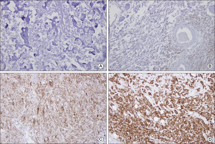
Representative areas, without spontaneous necrosis, were selected from incisional biopsy samples. We used a Zymed nonbiotin amplification system (Zymed Laboratories, South San Francisco, CA). Primary goat anti-human EGFR polyclonal anti-body and anti-goat EGFR biotinylated secondary antibody were purchased from Santa Cruz Biotechnology (Santa Cruz, CA). EGFR immunoreactivity was observed in membranes or cytoplasm of osteosarcoma cells. Some tissues showed strong immunoreactivity for EGFR, whereas others showed moderate or no immunoreactivity. Staining intensity was interpreted as follows: low, positivity of <10% of tumor cells; intermediate, positivity of 10-50% of tumor cells; and high, positivity of >50% of tumor cells.
3. Cell proliferation assays
Human osteosarcoma cell lines HOS, KHOS/NP, MG-63, and U-2 OS were obtained from the American Type Culture Collection (ATCC; Manassas, VA). Gefitinib and BIBW2992 were purchased from SelleckChem (S1025, S1011, Houston, TX), dissolved in dimethyl sulfoxide, and stored at -20℃. In each well of 96-well plates, 5,000 cells in 50 µL of media were seeded and incubated overnight at 37℃ in a humidified 5% CO2 incubator. On the following day, gefitinib and BIBW 2992 (0.01-20 µM) were added to wells and plates, followed by incubation for an additional 72 hours. A 3-(4, 5-dimethylthiazol-2-yl)-2, 5-diphenyltetrazolium bromide (MTT) assay was used for measurement of cell proliferation. IC50 values were determined from dose-response curves.
4. Western blot analysis
Cell lysates were prepared using a kit from Cell Signaling Technology (Beverly, MA). Equal amounts of protein (determined using the Bio-Rad Protein assay, Bio-Rad, Hercules, CA) were run on 12% sodium dodecyl sulfate polyacrylamide gel electrophoresis gels and transferred to polyvinylidene fluoride membranes, which were washed with Tris-buffered saline (TBS) containing 0.1% Tween 20 (TTBS), blocked with TTBS and 5% wt/vol nonfat dry milk overnight at 4℃, and incubated with primary antibody at 1 : 1,000 dilution. Membranes were then washed with TTBS, followed by incubation with horseradish peroxidase-conjugated secondary antibody (1 : 1,000). Signals were detected using an Amersham ECL (GE Healthcare Life Sciences, Munich, Germany).
5. EGFR mutation analysis
The genomic DNAs of the four osteosarcoma cell lines were extracted using a High Pure PCR template Preparation kit (Roche, Mannheim, Germany). PCR and real-time amplification monitoring of peptide nucleic acid PCR clamping were performed using a CFX 96 system (Bio-Rad). Genomic PCR was performed in final volume DNA and 0.75 U Taq polymerase (Solgent, Daejeon, Korea). The following sequencing primers were employed: E18-SF, 5-AGGTGACCCTTGTCTCTGTG-3, E18-SR, and 5-CCTGTGCCAGGGACCTTAC-3 for exon 18; E19-SF, 5-CATGTGGCACCATCTCACAA-3, E19-SR, and 5-CCCACA CAGCAAAGCAGAA-3 for exon 19; E20-B-F, 5-ATCGCATTCATGCGTCTTC-3, E20-B-R, and 5-GTCTTTGTGTTCCCGGACAT-3 for exon 20; E21-B-F, 5-CCTCACAGCAGGGTCTTCTC-3, E21-BR, and 5-GGAAAATGCTGGCTGACCTA-3 for exon 21. Sequences were compared with the GenBank-archived sequence of human EGFR.
6. Gene expression analysis
A Low Input Quick Amp labeling kit (Agilent Technologies, Santa Clara, CA) was used for labeling and amplification of total RNA samples (200 ηg), and Cy3-labeled amplified RNAs (aRNA) were re-suspended in 50 µL of hybridization solution (Agilent Technologies). Labeled aRNAs were then placed on an Agilent SurePrint G3 Human GE 8×60K array (Agilent Technologies) and covered with a Gasket 8-plex slide (Agilent Technologies). The slides were hybridized for 17 hours at 65℃, washed in 2× saline sodium citrate (SSC) containing 0.1% sodium dodecyl sulfate for 2 minutes, 1×SSC for 3 minutes, and then in 0.2×SSC for 2 minutes at room temperature, followed by centrifugation at 1,800× g for 20 seconds for drying. An Agilent scanner and its on-board software were used for analysis of arrays. Feature Extraction v10.7.3.1 (Agilent Technologies) was used for calculation of gene expression levels, and the Robust Multi-Array Average algorithm was used for generation of relative signal intensities for each gene. The quantile normalization method in GeneSpring GX 11.5.1 (Agilent Technologies) was used for processing of data.
7. Statistical analysis
The chi-square test was used for analysis of correlations between categorical clinicopathologic variables and EGFR expression. Variables analyzed included age, sex, tumor location, histologic subtype, tumor volume, and histologic response to preoperative chemotherapy. Tumor volumes were calculated using the ellipsoid formula [V = (4π/3)abc, where a, b, and c represent length, width, and depth] [19]. Kaplan-Meier survival analysis was used to examine relations between clinicopathologic variables and events (defined as either local recurrence, metastasis, or death). The log-rank test was used for calculation of survival differences according to clinicopathologic variables. SPSS ver. 11.5 (SPSS Inc., Chicago, IL) was used in performance of all calculations, and p-values of <0.05 were considered significant.
1. EGFR protein expression, clinicopathologic characteristics, and outcome
Twenty-seven (90.0%) biopsy samples showed positive staining for EGFR protein (Table 1, Fig. 1); staining intensities for EGFR protein were as follows: high in 9, intermediate in 12, and low in 6. Neither the presence of EGFR staining nor its intensity showed an association with age, sex, tumor location, or histologic response to preoperative chemotherapy (Table 2). However, intermediate or high staining intensity for EGFR was related to a smaller tumor volume (p<0.001) and a histologic subtype other than osteoblastic type (p=0.03).
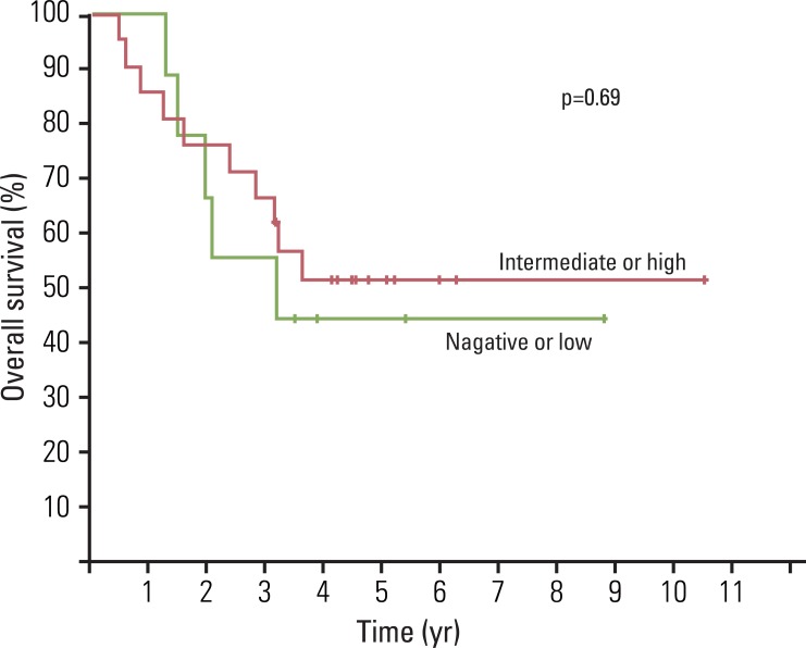
In addition, EGFR positivity and its staining intensity did not appear to be related to the survival (Fig. 2). Five-year OS for our 30 patients was 49.2±9.3%. Patients were followed for a median of 40.4 months (range, 6.0 to 126.0 months), and there were 17 events (14 distant metastases, 1 local recurrence, and 2 local recurrences combined with distant metastasis). Metastatic sites were as follows: lung (n=9), bone (n=3), lung and bone (n=3), adrenal gland (n=1), and pancreas (n=1). Median event-free survival for the 17 patients who experienced an adverse event was 9.3 months (range, 2.0 to 40.4 months).
2. Effect of EGFR inhibitors on the viabilities of osteosarcoma cell lines
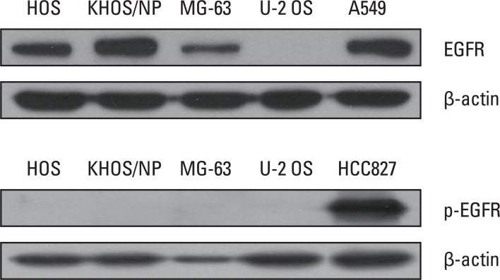
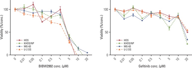
For the four osteosarcoma cell lines tested, expression of EGFR protein was observed in HOS, MG-63, and KHOS/NP cells, however, expression of p-EGFR was not observed in any cell line under basal culture conditions (Fig. 3). Osteosarcoma cells were exposed to gefitinib or BIBW2992 at concentrations ranging from 0 to 20 µM for 72 hours. However, no dose-dependent inhibitory effect was observed in any of the cell lines (Fig. 4).
3. Gene expression and mutation analysis of osteosarcoma cell lines
Table 3 shows expression of EGFR and phosphatase and tensin homolog (PTEN) in osteosarcoma cell lines. Expression of EGFR was observed at higher levels and its level of expression showed concurrence with western blot findings. However, PTEN was expressed at lower levels.
In the meantime, the tyrosine kinase domain from exons 18 to 21 was sequenced in all four cell lines, in order to determine whether they harbor constitutively activating mutations that confer sensitivity to tyrosine kinase inhibitors. We found single base changes at codon 2361 in exon 20 in HOS, MG-63, and KHOS/NP cells (G to A). However, these changes did not result in amino acid changes. Furthermore, we failed to find any known mutation associated with constitutive activation of EGFR in any of the four cell lines.
In the present study, we found that EGFR protein is frequently expressed in osteosarcoma biopsy samples and cell lines. However, we found no association between expression of EGFR protein and treatment outcome, and, in addition, EGFR inhibitors gefitinib and BIBW2992 had no significant inhibitory effect on the viabilities of osteosarcoma cells. A single nucleotide change in the EGFR tyrosine kinase domain was identified by mutation analysis in all four osteosarcoma cell lines examined, however, no activating mutation was found.
Some limitations of the present study must be considered. First, due to the retrospective nature of this study, our study cohort included only a small number of patients who were treated over two decades. Thus, patients were not prospectively randomized and treatments were heterogeneous. Second, we believe that selection bias must have existed; for example, inclusion of patients who are older than 40 years. And the poor survival might have masked the prognostic significance of EGFR expression. Third, the reliance on immunohistochemistry and semiquantitative measures used for assessment of EGFR expression imposes limitations. Forth, although EGFR mutation status should have been determined in biopsy samples, due to the small sample sizes, it was limited to the four cell lines.
Relations between EGFR expression and the clinicopathologic characteristics of osteosarcoma remain elusive. In previous studies, involvement of EGFR in progression of lung, colon, head, and neck cancers has been suggested [7-11,20]. Thus, we supposed that EGFR expression might be related to the aggressive characteristics and poor treatment outcome of osteosarcoma. However, we found no relation between EGFR expression and treatment outcome; actually, an association with a small tumor volume was observed, which was not expected. We believe that this finding was probably due to the small sample size and section bias, however, it may be that the role of EGFR in osteosarcoma differs from that in adult carcinomas. Little data are available on the association between EGFR expression and clinical outcome in osteosarcoma. Freeman et al. [16] also failed to detect any association between EGFR expression and clinical findings in osteosarcoma, however, Kersting et al. [18] concluded that EGFR expression has a dose-dependent association with better prognosis in patients with high-grade osteosarcoma. Conduct of additional studies to elucidate the mechanism responsible for EGFR expression and for the clinicopathologic characteristics of osteosarcoma will be needed.
Contrary to our expectation, we observed no inhibitory effect of gefitinib and BIBW2992 on the viabilities of osteosarcoma cells. In order for EGFR-directed therapy to be available in osteosarcoma, assessment of its activation status and impact of its signaling on cell proliferation as well as the relationship between its expression and outcome need to be clarified. It has previously been suggested that osteosarcoma might be biologically dependent on EGFR-modulated cell signaling pathways [12,13,15,21,22]. Although expression of EGFR protein was observed in the four osteosarcoma cell lines that we tested, none of them expressed p-EGFR. It has been suggested that neither the expression level nor the phosphorylation status of EGFR showed correlation with sensitivity to gefitinib [23]. Although we tested gefitinib and BIBW2992 at different concentrations, we failed to observe any significant inhibitory effect on cell viability. The mutational status of EGFR has been reported to modulate responsiveness to EGFR inhibitors [9,24], therefore, we analyzed the tyrosine kinase domain of EGFR in the four osteosarcoma cell lines. Although an EGFR polymorphism was found in exon 20, it did not result in an amino acid change. In addition, we did not identify any activating mutation known to confer susceptibility to EGFR inhibitors. The osteosarcoma cell lines used in the present study exhibited slightly lower PTEN expression, loss of which is associated with resistance to anti-EGFR tyrosine kinase inhibitors [9,24]. The possibility exists that the lack of EGFR mutation and efficacy of EGFR inhibitors might be related to the cells we used. The cell lines analyzed were not of early passages, but were of multiple passages that might have accumulated many changes that were not present in the primary tumor specimens. Or, other EGFR-directed treatment modalities might be effective in osteosarcoma. Recently it was reported that anti-EGFR antibody cetuximab enhances the cytolytic activity of natural killer cell toward osteosarcoma cells [25]. Available data suggest the probable existence of subtle interplay between different growth factor receptors and downstream molecules in osteosarcoma. Conduct of further studies to elucidate the roles of EGFR and its inhibitors in osteosarcoma will be needed.
We found that EGFR protein is frequently expressed in osteosarcoma, however, this expression showed no association with treatment outcome. In addition, the two EGFR inhibitors, gefitinib and BIBW2992, had no observable inhibitory effect on viability of osteosarcoma cells, and mutation analysis of the EGFR tyrosine kinase domain failed to identify any activating mutation. However, considering that our study was performed using a limited number of biopsy samples and cell lines, and evaluated two kinds of EGFR inhibitors, conduct of further studies to explore the potential of other therapeutic agents targeting EGFR will be necessary.
Acknowledgements
This study was supported by the Clinical Radiological Research Project, Korea Institute of Radiological and Medical Sciences (Grant No. 50459-2010).

Conflicts of interest relevant to this article was not reported.

  • 1. Bacci G, Longhi A, Fagioli F, Briccoli A, Versari M, Picci P. Adjuvant and neoadjuvant chemotherapy for osteosarcoma of the extremities: 27 year experience at Rizzoli Institute, Italy. Eur J Cancer. 2005;41:2836–2845. PMID: 16298125ArticlePubMed
  • 2. Gorlick R, Anderson P, Andrulis I, Arndt C, Beardsley GP, Bernstein M, et al. Biology of childhood osteogenic sarcoma and potential targets for therapeutic development: meeting summary. Clin Cancer Res. 2003;9:5442–5453. PMID: 14654523PubMed
  • 3. Bacci G, Forni C, Ferrari S, Longhi A, Bertoni F, Mercuri M, et al. Neoadjuvant chemotherapy for osteosarcoma of the extremity: intensification of preoperative treatment does not increase the rate of good histologic response to the primary tumor or improve the final outcome. J Pediatr Hematol Oncol. 2003;25:845–853. PMID: 14608193ArticlePubMed
  • 4. Eselgrim M, Grunert H, Kuhne T, Zoubek A, Kevric M, Burger H, et al. Dose intensity of chemotherapy for osteosarcoma and outcome in the Cooperative Osteosarcoma Study Group (COSS) trials. Pediatr Blood Cancer. 2006;47:42–50. PMID: 16206218ArticlePubMed
  • 5. Kager L, Zoubek A, Potschger U, Kastner U, Flege S, Kempf-Bielack B, et al. Primary metastatic osteosarcoma: presentation and outcome of patients treated on neoadjuvant Cooperative Osteosarcoma Study Group protocols. J Clin Oncol. 2003;21:2011–2018. PMID: 12743156ArticlePubMed
  • 6. Meyers PA, Gorlick R, Heller G, Casper E, Lane J, Huvos AG, et al. Intensification of preoperative chemotherapy for osteogenic sarcoma: results of the Memorial Sloan-Kettering (T12) protocol. J Clin Oncol. 1998;16:2452–2458. PMID: 9667263ArticlePubMed
  • 7. Herbst RS, Kies MS. ZD1839 (Iressa) in non-small cell lung cancer. Oncologist. 2002;7(Suppl 4):9–15. PMID: 12202783Article
  • 8. Raben D, Helfrich BA, Chan D, Johnson G, Bunn PA Jr. ZD1839, a selective epidermal growth factor receptor tyrosine kinase inhibitor, alone and in combination with radiation and chemotherapy as a new therapeutic strategy in non-small cell lung cancer. Semin Oncol. 2002;29(1 Suppl 4):37–46. PMID: 11894012ArticlePubMed
  • 9. Vivanco I, Mellinghoff IK. Epidermal growth factor receptor inhibitors in oncology. Curr Opin Oncol. 2010;22:573–578. PMID: 20739887ArticlePubMed
  • 10. Yarom N, Jonker DJ. The role of the epidermal growth factor receptor in the mechanism and treatment of colorectal cancer. Discov Med. 2011;11:95–105. PMID: 21356164PubMed
  • 11. Sharafinski ME, Ferris RL, Ferrone S, Grandis JR. Epidermal growth factor receptor targeted therapy of squamous cell carcinoma of the head and neck. Head Neck. 2010;32:1412–1421. PMID: 20848399ArticlePubMedPMC
  • 12. Dobashi Y, Suzuki S, Sugawara H, Ooi A. Involvement of epidermal growth factor receptor and downstream molecules in bone and soft tissue tumors. Hum Pathol. 2007;38:914–925. PMID: 17376509ArticlePubMed
  • 13. Dobashi Y, Takei N, Suzuki S, Yoneyama H, Hanawa M, Ooi A. Aberration of epidermal growth factor receptor expression in bone and soft-tissue tumors: protein overexpression, gene amplification and activation of downstream molecules. Mod Pathol. 2004;17:1497–1505. PMID: 15254554ArticlePubMed
  • 14. Oda Y, Wehrmann B, Radig K, Walter H, Rose I, Neumann W, et al. Expression of growth factors and their receptors in human osteosarcomas Immunohistochemical detection of epidermal growth factor, platelet-derived growth factor and their receptors: its correlation with proliferating activities and p53 expression. Gen Diagn Pathol. 1995;141:97–103. PMID: 8548600PubMed
  • 15. Do SI, Jung WW, Kim HS, Park YK. The expression of epidermal growth factor receptor and its downstream signaling molecules in osteosarcoma. Int J Oncol. 2009;34:797–803. PMID: 19212684PubMed
  • 16. Freeman SS, Allen SW, Ganti R, Wu J, Ma J, Su X, et al. Copy number gains in EGFR and copy number losses in PTEN are common events in osteosarcoma tumors. Cancer. 2008;113:1453–1461. PMID: 18704985ArticlePubMedPMC
  • 17. Wen YH, Koeppen H, Garcia R, Chiriboga L, Tarlow BD, Peters BA, et al. Epidermal growth factor receptor in osteosarcoma: expression and mutational analysis. Hum Pathol. 2007;38:1184–1191. PMID: 17509661ArticlePubMed
  • 18. Kersting C, Gebert C, Agelopoulos K, Schmidt H, van Diest PJ, Juergens H, et al. Epidermal growth factor receptor expression in high-grade osteosarcomas is associated with a good clinical outcome. Clin Cancer Res. 2007;13:2998–3005. PMID: 17505002ArticlePubMed
  • 19. Lee JA, Kim MS, Kim DH, Lim JS, Yoo JY, Koh JS, et al. Relative tumor burden predicts metastasis-free survival in pediatric osteosarcoma. Pediatr Blood Cancer. 2008;50:195–200. PMID: 18061933ArticlePubMed
  • 20. Batus M, Fidler MJ, Bonomi PD. Primary and secondary therapeutic strategies for EGF receptor pathway inhibition in non-small-cell lung cancer. Expert Rev Anticancer Ther. 2010;10:1589–1599. PMID: 20942630ArticlePubMed
  • 21. Hughes DP, Thomas DG, Giordano TJ, McDonagh KT, Baker LH. Essential erbB family phosphorylation in osteosarcoma as a target for CI-1033 inhibition. Pediatr Blood Cancer. 2006;46:614–623. PMID: 16007579ArticlePubMed
  • 22. Stewart CF, Leggas M, Schuetz JD, Panetta JC, Cheshire PJ, Peterson J, et al. Gefitinib enhances the antitumor activity and oral bioavailability of irinotecan in mice. Cancer Res. 2004;64:7491–7499. PMID: 15492275ArticlePubMed
  • 23. Sirotnak FM, Zakowski MF, Miller VA, Scher HI, Kris MG. Efficacy of cytotoxic agents against human tumor xenografts is markedly enhanced by coadministration of ZD1839 (Iressa), an inhibitor of EGFR tyrosine kinase. Clin Cancer Res. 2000;6:4885–4892. PMID: 11156248PubMed
  • 24. Mellinghoff IK, Wang MY, Vivanco I, Haas-Kogan DA, Zhu S, Dia EQ, et al. Molecular determinants of the response of glioblastomas to EGFR kinase inhibitors. N Engl J Med. 2005;353:2012–2024. PMID: 16282176ArticlePubMed
  • 25. Pahl JH, Ruslan SE, Buddingh EP, Santos SJ, Szuhai K, Serra M, et al. Anti-EGFR antibody cetuximab enhances the cytolytic activity of natural killer cells toward osteosarcoma. Clin Cancer Res. 2012;18:432–441. PMID: 22090361ArticlePubMed
Fig. 1
Immunohistochemical staining for epidermal growth factor receptor protein in initial biopsy samples from patients with osteosarcoma. (A) Negative. (B) Low. (C) Intermediate. (D) High (A-D, ×400).
crt-44-202-g001.jpg
Fig. 2
Epidermal growth factor receptor protein expression and survival of patients with localized osteosarcoma.
crt-44-202-g002.jpg
Fig. 3
Epidermal growth factor receptor (EGFR) and p-EGFR expression of osteosarcoma cell lines in basal culture conditions.
crt-44-202-g003.jpg
Fig. 4
Effect of gefitinib and BIBW2992 on viability of osteosarcoma cells.
crt-44-202-g004.jpg
Table 1
Patients' Characteristics
Case No. Age (yr)/
Gender
Location Tumor
volume (mL)
Histologic
response
EGFR protein
expression
Recurrence
(mo)
Metastasis
(mo)
Follow-up
(mo)
Final
status
1 5/M Humerus, proximal 28.6 50 High - 28.1 38.0 DOD
2 9/F Femur, distal 127.2 100 High - - 71.4 CDF
3 12/F Femur, distal 1,335.6 80 Intermediate - 25.6 38.7 DOD
4 12/M Tibia, proximal 26.0 80 High - - 53.9 CDF
5 13/M Pelvis 344.5 90 Low 13.5 17.5 23.4 DOD
6 13/M Femur, distal 19.1 50 Intermediate - - 49.6 CDF
7 14/M Humerus, proximal 159.0 70 Negative - 22.3 38.3 DOD
8 14/M Tibia, proximal 21.5 10 High - - 50.6 CDF
9 15/F Femur, distal 333.9 95 Low - - 42.1 CDF
10 16/F Femur, distal 1,026.1 50 Low 8.6 3.6 24.8 DOD
11 18/M Tibia, proximal 19.1 100 High - 75.1 CDF
12 19/M Femur, distal 229.0 95 Intermediate - 57.2 CDF
13 20/M Femur, distal 44.5 NA Intermediate 29.5 29.1 33.9 DOD
14 22/M Tibia, proximal 48.7 30 High - - 126.0 CDF
15 22/M Humerus, proximal 55.7 10 Intermediate - 6.3 28.9 DOD
16 24/M Femur, distal 107.3 50 Intermediate - 40.4 62.5 NED
17 25/M Femur, proximal 62.0 60 Intermediate 7.6 7.6 14.8 DOD
18 27/F Femur, distal 67.8 0 Low - - 46.8 CDF
19 29/F Tibia, proximal 79.5 10 Negative - - 64.8 CDF
20 37/M Femur, distal 76.3 30 Intermediate - - 54.5 CDF
21 41/F Femur, distal 111.3 NA High - 3.1 19.1 DOD
22 42/F Humerus, shaft 172.3 50 Low - 12.6 105.3 NED
23 43/F Clavicle 31.0 99 Intermediate - - 60.6 CDF
24 47/M Femur, distal 59.4 50 High - - 38.2 CDF
25 49/M Humerus, proximal 198.8 75 Low - 9.0 15.7 DOD
26 57/F Tibia, proximal 174.9 30 Intermediate - 31.0 43.5 DOD
27 60/F Femur, distal 136.3 50 Intermediate - 5.7 10.4 DOD
28 62/M Rib 9.5 50 Intermediate - 3.7 7.1 DOD
29 71/F Tibia, proximal 152.6 NA Negative - 9.3 18.0 DOD
30 79/F Tibia, distal 19.1 NA high - 2.0 6.0 DOD

EGFR, epidermal growth factor receptor; M, male; DOD, died of disease; F, female; CDF, continuously disease-free; NA, not available; NED, no evidence of disease.

Table 2
Clinicopathologic variables and EGFR protein expression
Clinical variables No. of cases Cases with intermediate or high
expression for EGFR protein (%)
p-value
Age (yr) <40 20 14 (70.0) 1.00
≥40 10 7 (70.0)
Gender Male 17 14 (82.4) 0.09
Female 13 7 (53.8)
Tumor location Distal femur 13 10 (76.9) 0.26
Proximal tibia 7 5 (71.4)
Proximal Humerus 5 2 (40.0)
Other extremities 2 2 (100)
Pelvis 1 0 (0)
Elsewhere 2 5 (100)
Tumor volume (mL) <150 20 18 (90.0) 0.001
≥150 10 3 (30.0)
Histologic subtype Osteoblastic 13 5 (38.5) 0.03
Chondroblastic 1 1 (100)
Fibroblastic 9 8 (88.9)
Others 4 4 (100)
Histologic response Good 6 4 (66.7) 0.97
Poor 21 15 (71.4)

EGFR, epidermal growth factor receptor.

Table 3
Up- or down-regulation of genes in osteosarcoma cells vs. universal human reference RNA
Gene symbol OS cell Mean (A) Mean (B) Fold change (A/B)
EGFR HOS 167.70 79.35 2.11
KHOS/NP 159.82 79.35 2.01
MG-63 105.68 79.35 1.33
U-2 OS 89.69 79.35 1.13
PTEN HOS 1,109.08 1,241.50 0.89
KHOS/NP 1,291.89 1,241.50 1.04
MG-63 970.81 1,241.50 0.78
U-2 OS 900.12 1,241.50 0.72

Genes are ranked in order of fold change and are listed with their gene symbol ID, mean osteosarcoma cells (mean A) and universal human reference RNA (mean B), and with their gene description. EGFR, epidermal growth factor receptor; PTEN, phosphatase and tensin homolog.

Figure & Data

REFERENCES

    Citations

    Citations to this article as recorded by  
    • Combination Treatment with Free Doxorubicin and Inductive Moderate Hyperthermia for Sarcoma Saos-2 Cells
      Valerii E. Orel, Anatolii G. Diedkov, Vasyl V. Ostafiichuk, Sergii A. Lyalkin, Igor O. Tkachenko, Denys L. Kolesnyk, Valerii B. Orel, Olga Yo. Dasyukevich, Oleksandr Yu. Rykhalskyi, Oleksii V. Movchan, Alexander Yu. Galkin, Anna B. Prosvietova
      Pharmaceuticals.2025; 18(6): 852.     CrossRef
    • Urolithin A suppressed osteosarcoma cell migration and invasion via targeting MMPs and AKT1
      Abdolreza Ahmadi, Fatemehsadat Hosseini, Milad Iranshahy, Fatemeh B. Rassouli
      Scientific Reports.2025;[Epub]     CrossRef
    • PD-L1, STAT3, IL6, and EGFR Immunoexpressions in High-Grade Osteosarcoma
      Nayla Rahmadiani, Eviana Norahmawati, Agustina Tri Endharti, Ailen Oktaviana Hambalie, Satria Pandu Persada Isma, Raffaele Vitiello
      Advances in Orthopedics.2024; 2024: 1.     CrossRef
    • Optimizing near infrared laser irradiation and photosensitizer accumulation period for indocyanine green-mediated photodynamic therapy in breast cancer xenografts: a focus on treatment and characterization
      Hasim Ozgur Tabakoglu, Tuğba Kiriş Aydoğan, Ayşenur Kiriş, Saadet Akbulut
      Lasers in Medical Science.2024;[Epub]     CrossRef
    • EGFR-targeting peptide conjugated polymer–lipid hybrid nanoparticles for delivery of salinomycin to osteosarcoma
      Longhai Du, Yanlong Xu, Binxu Han, Yu Wang, Qingmin Zeng, Minghao Shao, Zuochong Yu
      Journal of Cancer Research and Therapeutics.2023; 19(6): 1544.     CrossRef
    • Epidermal growth factor signalling pathway in endochondral ossification: an evidence-based narrative review
      L. Mangiavini, G. M. Peretti, B. Canciani, N. Maffulli
      Annals of Medicine.2022; 54(1): 37.     CrossRef
    • Synergistic effect of growth factor receptor-bound protein 2/epidermal growth factor receptor dual-targeting peptide inhibitor and salinomycin on osteosarcoma
      Zuochong Yu, Yanlong Xu, Longhai Du, Fan Zhang, Minghao Shao, Lin Xie, Guoping Cai, Feizhou Lyu
      Journal of Cancer Research and Therapeutics.2022; 18(2): 352.     CrossRef
    • Stattic sensitizes osteosarcoma cells to epidermal growth factor receptor inhibitors via blocking the interleukin 6-induced STAT3 pathway
      Shenglin Wang, Yunqing Wang, Zhen Huang, Hongxiang Wei, Xinwen Wang, Rongkai Shen, Wenbin Lan, Guangxian Zhong, Jianhua Lin
      Acta Biochimica et Biophysica Sinica.2021; 53(12): 1670.     CrossRef
    • Afatinib is active in osteosarcoma in osteosarcoma cell lines
      Marlid Cruz-Ramos, Yessica Zamudio-Cuevas, Daniel Medina-Luna, Karina Martínez-Flores, Gabriela Martínez-Nava, Javier Fernández-Torres, Alberto López-Reyes, Flavio Solca
      Journal of Cancer Research and Clinical Oncology.2020; 146(7): 1693.     CrossRef
    • The synergistic inhibitory effect of combining therapies targeting EGFR and mitochondria in sarcomas
      Xiaochun Wang, Reichelle X. Yeo, Philip J. Hogg, David Goldstein, Philip Crowe, Pierre J. Dilda, Jia-Lin Yang
      Oncotarget.2020; 11(1): 46.     CrossRef
    • Targeting Molecular Mechanisms Underlying Treatment Efficacy and Resistance in Osteosarcoma: A Review of Current and Future Strategies
      Ingrid Lilienthal, Nikolas Herold
      International Journal of Molecular Sciences.2020; 21(18): 6885.     CrossRef
    • Prospects for NK Cell Therapy of Sarcoma
      Mieszko Lachota, Marianna Vincenti, Magdalena Winiarska, Kjetil Boye, Radosław Zagożdżon, Karl-Johan Malmberg
      Cancers.2020; 12(12): 3719.     CrossRef
    • Advanced development of ErbB family-targeted therapies in osteosarcoma treatment
      Wei Wang, Hua-fu Zhao, Teng-fei Yao, Hao Gong
      Investigational New Drugs.2019; 37(1): 175.     CrossRef
    • The TGFβ-miR-499a-SHKBP1 pathway induces resistance to EGFR inhibitors in osteosarcoma cancer stem cell-like cells
      Tian Wang, Dexing Wang, Lian Zhang, Ping Yang, Jing Wang, Qi Liu, Fei Yan, Feng Lin
      Journal of Experimental & Clinical Cancer Research.2019;[Epub]     CrossRef
    • Interpretation of Tongue Squamous Cell Carcinoma via Protein-Protein Interaction Network Construction and Analysis
      Mona Zamanian Azodi, Mostafa Rezaei-Tavirani, Majid Rezaei-Tavirani, Vahid Mansouri, Reza Vafaee
      International Journal of Cancer Management.2018;[Epub]     CrossRef
    • EGFR is required for FOS‐dependent bone tumor development via RSK2/CREB signaling
      Markus Linder, Elisabeth Glitzner, Sriram Srivatsa, Latifa Bakiri, Kazuhiko Matsuoka, Parastoo Shahrouzi, Monika Dumanic, Philipp Novoszel, Thomas Mohr, Oliver Langer, Thomas Wanek, Markus Mitterhauser, Erwin F Wagner, Maria Sibilia
      EMBO Molecular Medicine.2018;[Epub]     CrossRef
    • Prognostic significance of the expression of HER family members in primary osteosarcoma
      Sheng‑Lin Wang, Guang‑Xian Zhong, Xin‑Wen Wang, Feng‑Qiang Yu, Dan‑Feng Weng, Xin‑Xing Wang, Jian‑Hua Lin
      Oncology Letters.2018;[Epub]     CrossRef
    • Effects of epidermal growth factor receptor kinase inhibition on radiation response in canine osteosarcoma cells
      Fernanda B. Mantovani, Jodi A. Morrison, Anthony J. Mutsaers
      BMC Veterinary Research.2016;[Epub]     CrossRef
    • Evaluation of CD146 as Target for Radioimmunotherapy against Osteosarcoma
      Sara Westrøm, Tina B. Bønsdorff, Nasir Abbas, Øyvind S. Bruland, Thora J. Jonasdottir, Gunhild M. Mælandsmo, Roy H. Larsen, Salvatore V Pizzo
      PLOS ONE.2016; 11(10): e0165382.     CrossRef
    • Osteoblast dysfunctions in bone diseases: from cellular and molecular mechanisms to therapeutic strategies
      Pierre J. Marie
      Cellular and Molecular Life Sciences.2015; 72(7): 1347.     CrossRef
    • Receptor tyrosine kinases: Characterisation, mechanism of action and therapeutic interests for bone cancers
      Aude I. Ségaliny, Marta Tellez-Gabriel, Marie-Françoise Heymann, Dominique Heymann
      Journal of Bone Oncology.2015; 4(1): 1.     CrossRef
    • EGFR is not a major driver for osteosarcoma cell growth in vitro but contributes to starvation and chemotherapy resistance
      Florian Sevelda, Lisa Mayr, Bernd Kubista, Daniela Lötsch, Sushilla van Schoonhoven, Reinhard Windhager, Christine Pirker, Michael Micksche, Walter Berger
      Journal of Experimental & Clinical Cancer Research.2015;[Epub]     CrossRef
    • Amphiregulin enhances intercellular adhesion molecule-1 expression and promotes tumor metastasis in human osteosarcoma
      Ju-Fang Liu, Ya-Ting Tsao, Chun-Han Hou
      Oncotarget.2015; 6(38): 40880.     CrossRef
    • ZD6474, a new treatment strategy for human osteosarcoma, and its potential synergistic effect with celecoxib
      Jiani Liu, Jiangxue Wu, Ling Zhou, Changchuan Pan, Yi Zhou, Wuying Du, Jie-min Chen, Xiaofeng Zhu, Jingnan Shen, Shuai Chen, Ran-yi Liu, Wenlin Huang
      Oncotarget.2015; 6(25): 21341.     CrossRef
    • Molecular profiling of childhood cancer: Biomarkers and novel therapies
      Federica Saletta, Carol Wadham, David S. Ziegler, Glenn M. Marshall, Michelle Haber, Geoffrey McCowage, Murray D. Norris, Jennifer A. Byrne
      BBA Clinical.2014; 1: 59.     CrossRef
    • ErbB Receptors as Prognostic and Therapeutic Drug Targets in Bone and Soft Tissue Sarcomas
      Hongsheng Wang, Qingbo Yang, Zeze Fu, Dongqing Zuo, Yingqi Hua, Zhengdong Cai
      Cancer Investigation.2014; 32(10): 533.     CrossRef
    • Imatinib Mesylate Exerts Anti-Proliferative Effects on Osteosarcoma Cells and Inhibits the Tumour Growth in Immunocompetent Murine Models
      Bérengère Gobin, Gatien Moriceau, Benjamin Ory, Céline Charrier, Régis Brion, Frederic Blanchard, Françoise Redini, Dominique Heymann, Gerard Roel Rutteman
      PLoS ONE.2014; 9(3): e90795.     CrossRef
    • Focal Chromosomal Copy Number Aberrations Identify CMTM8 and GPR177 as New Candidate Driver Genes in Osteosarcoma
      Joeri Both, Oscar Krijgsman, Johannes Bras, Gerard R. Schaap, Frank Baas, Bauke Ylstra, Theo J. M. Hulsebos, David Loeb
      PLoS ONE.2014; 9(12): e115835.     CrossRef
    • Gefitinib and Luteolin Cause Growth Arrest of Human Prostate Cancer PC-3 Cells via Inhibition of Cyclin G-Associated Kinase and Induction of miR-630
      Minami A. Sakurai, Yuki Ozaki, Daisuke Okuzaki, Yoko Naito, Towa Sasakura, Ayumi Okamoto, Hiroe Tabara, Takao Inoue, Man Hagiyama, Akihiko Ito, Norikazu Yabuta, Hiroshi Nojima, Ichiro Aoki
      PLoS ONE.2014; 9(6): e100124.     CrossRef
    • Clinical Predictive Value of Serum Angiogenic Factor in Patients with Osteosarcoma
      Zhe Chen, Qi-Xin Chen, Zhao-Yang Hou, Jiong Hu, Yan-Guang Cao
      Asian Pacific Journal of Cancer Prevention.2012; 13(9): 4823.     CrossRef

    • PubReader PubReader
    • ePub LinkePub Link
    • Cite
      CITE
      export Copy Download
      Close
      Download Citation
      Download a citation file in RIS format that can be imported by all major citation management software, including EndNote, ProCite, RefWorks, and Reference Manager.

      Format:
      • RIS — For EndNote, ProCite, RefWorks, and most other reference management software
      • BibTeX — For JabRef, BibDesk, and other BibTeX-specific software
      Include:
      • Citation for the content below
      Epidermal Growth Factor Receptor: Is It a Feasible Target for the Treatment of Osteosarcoma?
      Cancer Res Treat. 2012;44(3):202-209.   Published online September 30, 2012
      Close
    • XML DownloadXML Download
    Epidermal Growth Factor Receptor: Is It a Feasible Target for the Treatment of Osteosarcoma?
    Image Image Image Image
    Fig. 1 Immunohistochemical staining for epidermal growth factor receptor protein in initial biopsy samples from patients with osteosarcoma. (A) Negative. (B) Low. (C) Intermediate. (D) High (A-D, ×400).
    Fig. 2 Epidermal growth factor receptor protein expression and survival of patients with localized osteosarcoma.
    Fig. 3 Epidermal growth factor receptor (EGFR) and p-EGFR expression of osteosarcoma cell lines in basal culture conditions.
    Fig. 4 Effect of gefitinib and BIBW2992 on viability of osteosarcoma cells.
    Epidermal Growth Factor Receptor: Is It a Feasible Target for the Treatment of Osteosarcoma?
    Case No. Age (yr)/
    Gender
    Location Tumor
    volume (mL)
    Histologic
    response
    EGFR protein
    expression
    Recurrence
    (mo)
    Metastasis
    (mo)
    Follow-up
    (mo)
    Final
    status
    1 5/M Humerus, proximal 28.6 50 High - 28.1 38.0 DOD
    2 9/F Femur, distal 127.2 100 High - - 71.4 CDF
    3 12/F Femur, distal 1,335.6 80 Intermediate - 25.6 38.7 DOD
    4 12/M Tibia, proximal 26.0 80 High - - 53.9 CDF
    5 13/M Pelvis 344.5 90 Low 13.5 17.5 23.4 DOD
    6 13/M Femur, distal 19.1 50 Intermediate - - 49.6 CDF
    7 14/M Humerus, proximal 159.0 70 Negative - 22.3 38.3 DOD
    8 14/M Tibia, proximal 21.5 10 High - - 50.6 CDF
    9 15/F Femur, distal 333.9 95 Low - - 42.1 CDF
    10 16/F Femur, distal 1,026.1 50 Low 8.6 3.6 24.8 DOD
    11 18/M Tibia, proximal 19.1 100 High - 75.1 CDF
    12 19/M Femur, distal 229.0 95 Intermediate - 57.2 CDF
    13 20/M Femur, distal 44.5 NA Intermediate 29.5 29.1 33.9 DOD
    14 22/M Tibia, proximal 48.7 30 High - - 126.0 CDF
    15 22/M Humerus, proximal 55.7 10 Intermediate - 6.3 28.9 DOD
    16 24/M Femur, distal 107.3 50 Intermediate - 40.4 62.5 NED
    17 25/M Femur, proximal 62.0 60 Intermediate 7.6 7.6 14.8 DOD
    18 27/F Femur, distal 67.8 0 Low - - 46.8 CDF
    19 29/F Tibia, proximal 79.5 10 Negative - - 64.8 CDF
    20 37/M Femur, distal 76.3 30 Intermediate - - 54.5 CDF
    21 41/F Femur, distal 111.3 NA High - 3.1 19.1 DOD
    22 42/F Humerus, shaft 172.3 50 Low - 12.6 105.3 NED
    23 43/F Clavicle 31.0 99 Intermediate - - 60.6 CDF
    24 47/M Femur, distal 59.4 50 High - - 38.2 CDF
    25 49/M Humerus, proximal 198.8 75 Low - 9.0 15.7 DOD
    26 57/F Tibia, proximal 174.9 30 Intermediate - 31.0 43.5 DOD
    27 60/F Femur, distal 136.3 50 Intermediate - 5.7 10.4 DOD
    28 62/M Rib 9.5 50 Intermediate - 3.7 7.1 DOD
    29 71/F Tibia, proximal 152.6 NA Negative - 9.3 18.0 DOD
    30 79/F Tibia, distal 19.1 NA high - 2.0 6.0 DOD
    Clinical variables No. of cases Cases with intermediate or high
    expression for EGFR protein (%)
    p-value
    Age (yr) <40 20 14 (70.0) 1.00
    ≥40 10 7 (70.0)
    Gender Male 17 14 (82.4) 0.09
    Female 13 7 (53.8)
    Tumor location Distal femur 13 10 (76.9) 0.26
    Proximal tibia 7 5 (71.4)
    Proximal Humerus 5 2 (40.0)
    Other extremities 2 2 (100)
    Pelvis 1 0 (0)
    Elsewhere 2 5 (100)
    Tumor volume (mL) <150 20 18 (90.0) 0.001
    ≥150 10 3 (30.0)
    Histologic subtype Osteoblastic 13 5 (38.5) 0.03
    Chondroblastic 1 1 (100)
    Fibroblastic 9 8 (88.9)
    Others 4 4 (100)
    Histologic response Good 6 4 (66.7) 0.97
    Poor 21 15 (71.4)
    Gene symbol OS cell Mean (A) Mean (B) Fold change (A/B)
    EGFR HOS 167.70 79.35 2.11
    KHOS/NP 159.82 79.35 2.01
    MG-63 105.68 79.35 1.33
    U-2 OS 89.69 79.35 1.13
    PTEN HOS 1,109.08 1,241.50 0.89
    KHOS/NP 1,291.89 1,241.50 1.04
    MG-63 970.81 1,241.50 0.78
    U-2 OS 900.12 1,241.50 0.72
    Table 1 Patients' Characteristics

    EGFR, epidermal growth factor receptor; M, male; DOD, died of disease; F, female; CDF, continuously disease-free; NA, not available; NED, no evidence of disease.

    Table 2 Clinicopathologic variables and EGFR protein expression

    EGFR, epidermal growth factor receptor.

    Table 3 Up- or down-regulation of genes in osteosarcoma cells vs. universal human reference RNA

    Genes are ranked in order of fold change and are listed with their gene symbol ID, mean osteosarcoma cells (mean A) and universal human reference RNA (mean B), and with their gene description. EGFR, epidermal growth factor receptor; PTEN, phosphatase and tensin homolog.


    Cancer Res Treat : Cancer Research and Treatment
    Close layer
    TOP