Skip Navigation
Skip to contents

Cancer Res Treat : Cancer Research and Treatment

OPEN ACCESS

Articles

Page Path
HOME > Cancer Res Treat > Volume 45(3); 2013 > Article
Original Article Prognostic Role of Interleukin-6, Interleukin-8, and Leptin Levels According to Breast Cancer Subtype
Young Ae Cho, PhD, MPH1, Mi-Kyung Sung, PhD2, Jee-Young Yeon, PhD2, Jungsil Ro, MD, PhD3, Jeongseon Kim, PhD1
Cancer Research and Treatment : Official Journal of Korean Cancer Association 2013;45(3):210-219.
DOI: https://doi.org/10.4143/crt.2013.45.3.210
Published online: September 30, 2013

1Molecular Epidemiology Branch, National Cancer Center, Goyang, Korea.

2Department of Food and Nutrition, Sookmyung Women's University, Seoul, Korea.

3Center for Breast Cancer, National Cancer Center, Goyang, Korea.

Correspondence: Jeongseon Kim, PhD. Molecular Epidemiology Branch, National Cancer Center, 111 Jungbalsanro, Ilsandong-gu, Goyang 410-769, Korea.
Tel: 82-31-920-2570, Fax: 82-31-920-2579, jskim@ncc.re.kr
• Received: October 10, 2012   • Accepted: April 3, 2013

Copyright © 2013 by the Korean Cancer Association

This is an Open-Access article distributed under the terms of the Creative Commons Attribution Non-Commercial License (http://creativecommons.org/licenses/by-nc/3.0/) which permits unrestricted non-commercial use, distribution, and reproduction in any medium, provided the original work is properly cited.

  • 13,521 Views
  • 128 Download
  • 34 Crossref
  • 47 Scopus
prev next
  • Purpose
    Inflammation within the tumor microenvironment has been reported to show an association with poor prognosis in breast cancer. However, the associations may differ according to breast cancer subtype. In this study, we investigated the association between inflammation-related markers and breast cancer recurrence according to patients' tumor subtypes.
  • Materials and Methods
    This prospective study included 240 patients who underwent surgery for management of newly diagnosed breast cancer. Levels of inflammation-related markers (interleukin [IL]-1β, IL-6, IL-8, monocyte chemoattractant protein-1 [MCP-1], leptin, and adiponectin) were measured at diagnosis, and the associations between these markers and breast cancer recurrence during a six-year follow-up period were examined using the Kaplan-Meier statistical method.
  • Results
    Overall, inflammation-related markers showed no association with breast cancer recurrence. However, when data were stratified by tumor subtype, higher levels of some mediators showed an association with poor prognosis among patients with particular subtypes. Compared to patients without recurrence, patients with recurrence had higher levels of circulating IL-6 (p=0.024) and IL-8 (p=0.016) only among those with HER2- tumors and had higher levels of leptin (p=0.034) only among those with estrogen receptor (ER)+/progesterone receptor (PR)+ tumors. Results of survival analyses revealed an association of high levels of IL-6 (p=0.016) and IL-8 (p=0.022) with poor recurrence-free survival in patients with HER2- tumors. In addition, higher leptin levels indicated shorter recurrence-free survival time only among patients with ER+/PR+ tumors (p=0.022).
  • Conclusion
    We found that certain cytokines could have a differential prognostic impact on breast cancer recurrence according to breast cancer subtype. Conduct of additional large studies will be required in order to elucidate the precise roles of these cytokines in breast cancer progression.
Correlation of inflammation within the tumor microenvironment with tumor growth, increased invasiveness, and poor prognosis in breast cancer has been reported [1]. The tumor microenvironment is rich in inflammation-related mediators, such as immune cell-derived cytokines, chemokines, and adipocyte-derived adipokines, which are secreted by either cancer cells or tumor-associated immune cells [1]. Many studies and reviews have demonstrated an association of increased levels of circulating interleukin (IL)-1β, IL-6, IL-8, monocyte chemoattractant protein-1 (MCP-1), or leptin with a poor prognosis in breast cancer patients [1,2]. It has been proposed that these mediators can stimulate proliferation and invasion of breast cancer cells directly or are involved in angiogenesis, which is essential for development and progression of breast cancer [3]. In contrast with these mediators, adiponectin, which shows negative correlation with leptin expression, is anti-angiogenic and anti-proliferative, and some studies have reported reduced adiponectin levels in breast cancer patients [2]. Therefore, these inflammation-related markers may be useful in prediction of prognosis and identification of cases with a high risk for metastasis [1,4].
Breast cancer is a complex and heterogeneous disease, and its prognosis may depend on characteristics of tumor and host [5]. Therefore, the roles of these inflammation-related markers in mediation of tumor growth and metastasis could be influenced by distinct subtypes that have been identified on the basis of gene or protein expression in tumor tissue [4]. Different breast cancer subtypes may produce distinct inflammatory mediators, which may affect their distinct tumor progression pathways [6,7]. The aim of the current study was to investigate the association between breast cancer recurrence and inflammation-related markers, including IL-1β, IL-6, IL-8, MCP-1, leptin, and adiponectin, according to patients' tumor subtype.
1. Study participants and follow-up
This was a prospective study of newly diagnosed breast cancer patients who underwent surgery at the National Cancer Center Hospital in Korea between July 2007 and September 2008, with follow-up through January 2013. Among 441 breast cancer patients, 26 patients did not agree to participate in the study and 105 patients refused to provide their blood for the study. Among 310 women, we excluded patients with a previous history of cancer (n=14) and those with stage 0 or IV cancer (n=56). The remaining 240 patients were included in the final analysis. Those patients were followed up for six years in order to identify cases of breast cancer recurrence. Breast cancer recurrence included local (n=8) or distant metastasis (n=23), and 31 recurrent patients were identified. Eleven recurrent patients died of breast cancer. Each participant provided written informed consent, and the procedure was approved by the Institutional Review Board of the National Cancer Center (IRB protocol number NCCNCS 07-083).
2. Data collection
Participants were interviewed in person by a trained researcher using a structured questionnaire. Data collected in baseline evaluations included demographic characteristics, personal and family medical history, alcohol consumption, smoking history, hormone replacement therapy, and age at menarche or menopause. Blood samples were collected at diagnosis of breast cancer and stored at -80℃ until analysis. The plasma concentrations of IL-1β, IL-6, IL-8, MCP-1, leptin, and adiponectin were quantified using the human Quantikine ELISA kit (R&D Systems, Minneapolis, MN) according to the manufacturer's instructions; absorbance was read on a plate reader (Biotech Instruments Inc., Winooski, VT).
3. Evaluation of breast cancer clinicopathological factors
We evaluated conventional clinicopathological factors, including adjuvant treatment modalities (hormone therapy and anti-HER2-therapy), tumor subtype, and Ki-67 index. Immunohistochemistry (IHC) of four different biological factors (estrogen receptor [ER], SP1, Ventana, Tucson, AZ; progesterone receptor [PR], 1E2, Ventana; HER2, polyclonal, Dako, Glostrup, Denmark; Ki-67, MIB-1, Dako) was performed using paraffin-embedded breast tumor sections according to reported recommendations for tumor marker prognostic studies (REMARK) [8]. ER and PR positivity was defined using a cut-off value of 10% or more of positively stained nuclei [9]. HER2 was scored as 0-3+ according to the method recommended for the Dako Hercep Test [10]. The HER2 status of each patient was defined as follows: HER2-positive (HER2+) if the IHC score was 3+, HER2-negative (HER2-) if the IHC score was 0 or 1+, or indeterminate if the IHC score was 2+. For indeterminate patient samples, further analysis was performed using fluorescence in situ hybridization (FISH); if FISH was not available, the patients were considered HER2+-unknown [11].
For assessment of Ki-67 in breast cancer, cells stained for Ki-67 were counted and expressed as a percentage. Ki-67 index of less than 15% was assessed as low expression [12]. The pathological tumor stage was assessed according to the criteria established by the seventh edition of the American Joint Committee on Cancer (AJCC) staging manual [13]. The tumor grade was determined according to the Scarff-Bloom-Richardson classification modified by Elston and Ellis [14].
4. Statistical analyses
All statistical analyses were performed using SAS ver. 9.1 (SAS Institute Inc., Cary, NC). A two-sided p-value of less than 0.05 was regarded as statistically significant.
For evaluation of differences in patients' clinocopatholgical characteristics (cancer stage, tumor subtype, tumor size, lymph node metastasis, Ki-67 index, histologic grade, and treatments) according to the levels of inflammation-related markers (IL-1β, IL-6, IL-8, MCP-1, leptin, and adiponectin), chi-square test and Kruskal Wallis test were used for categorical variables and continuous variables, respectively. Patients were divided into two groups (high/low) based on their median values of inflammation-related markers. In addition, patient characteristics (age, body mass index [BMI], smoking, alcohol intake, menopausal status, and clinicopathologic characteristics) were compared in relation to recurrence status using the Kaplan-Meier statistical method for estimation of recurrence-free survival, and the log-rank test for comparison of differences in recurrence-free survival. Recurrence-free survival was calculated from the day of sampling until breast cancer recurrence, death, or the end of the study period. Local and distant relapses were considered as recurrences. To investigate the association between the levels of inflammation-related markers and recurrence of breast cancer, the median levels of inflammation-related markers were compared according to patients' recurrence status; significant differences were identified using the median test. In addition, the Kaplan-Meier statistical method and the log-rank test were used for comparison of patients' recurrence-free survival rates according to the levels of inflammation-related markers. The Cox proportional hazards regression model was used to control for multiple factors simultaneously, and for estimation of the adjusted hazard ratios and the 95% confidence intervals. The following covariates were considered as potential confounders:
age, stage of disease, menopausal status, tumor subtypes, and tamoxifen treatment.
For all analyses, subgroup analyses were performed based on tumor subtype. Patients were divided into subgroups as follows: 1) four tumor subtypes: luminal A (ER+ and/or PR+, HER2-, and Ki-67 index<15%), luminal B ([ER+ and/or PR+, HER-, and Ki-67 index≥15%] or [ER+ and/or PR+, and HER2+]), HER2 only (ER-, PR-, and HER2+), and triple-negative (ER-, PR-, and HER2-); 2) ER/PR status: ER+/PR+ and ER-/PR-; 3) HER2 status: HER2+ and HER2-.
The median follow-up period was 57.9 months (interquartile range, 54.4 to 60.9 months) from the date of the initial breast cancer surgery. Based on tumor subtype, patients were divided into subgroups, as follows: in regard to the four tumor subtypes, 40.7% of patients had luminal A, 28.1% of patients had luminal B, 10.0% of patients had HER2+, and 21.3% of patients had triple negative tumors; in regard to ER/PR status, 71.3% of patients had ER+/PR+ tumors and 28.7% of patients had ER-/PR- tumors; in regard to HER2 status, 18.1% of patients had HER2+ tumors and 81.9% of patients had HER2- tumors.
Table 1 shows the different clinicopathological characteristics of patients according to the levels of inflammation-related markers (IL-1β, IL-6, IL-8, MCP-1, leptin, and adiponectin). No differences were observed with respect to breast cancer recurrence for all inflammation-related markers tested. However, differences in some clinicopathlogical prognostic factors were observed according to the levels of inflammation-related markers; IL-1β levels differed by tumor subtype (p=0.045); high IL-6 levels showed an association with advanced cancer stage (p=0.038) and lymph node metastasis (p=0.007); high MCP-1 levels showed an association with advanced cancer stage (p=0.025) and HER2+ tumors (p=0.016); high leptin levels showed an association with HER2+ tumors (p=0.016); high adiponectin levels showed an association with smaller tumor size (p=0.029).
Table 2 shows characteristics of patients according to recurrence status. No differences with respect to age, BMI, smoking status, alcohol intake, menopausal status, histologic grade, and treatments were observed between patients whose cancer recurred and those without recurrence. Patients who were in an advanced cancer stage (p=0.005), T stage (p=0.002) and N stage (p=0.002), and had higher Ki-67 index (p=0.013) had poor recurrence-free survival.
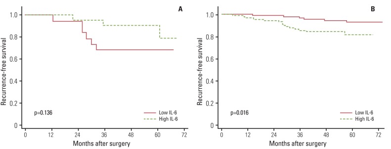
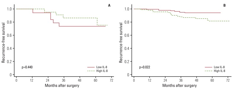
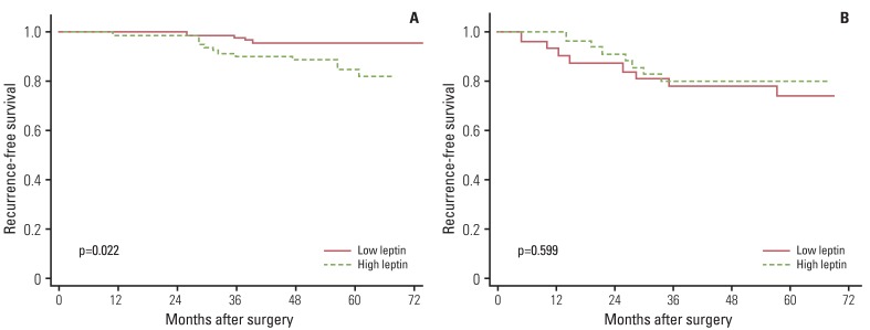
We examined the median plasma levels of inflammation-related markers (IL-1β, IL-6, IL-8, MCP-1, leptin, and adiponectin) according to patients' recurrence status, stratified by their tumor subtype. Overall, inflammation-related markers did not differ according to patients' recurrence status. However, different plasma levels of IL-6, IL-8, and leptin were observed according to patients' recurrence status only among patients with certain tumor subtypes (Table 3). Analysis of IL-6 levels according to patients' recurrence status showed higher levels of IL-6 in patients with triple negative (p=0.024) or HER2- (p=0.024) breast cancer with recurrence than in those without recurrence. Regarding IL-8, HER2- breast cancer patients with recurrence had higher IL-8 levels than those without recurrence (p=0.016). Regarding leptin, ER+/PR+ patients with recurrence had higher leptin levels than those without recurrence (p=0.034). However, the levels of IL-1β, MCP-1, and adiponectin did not differ according to patients' recurrence status in all subtypes tested (data not shown).
Finally, we compared recurrence-free survival according to the levels of inflammation-related markers. Overall, inflammation-related markers showed no association with recurrence-free survival. However, when data were stratified by breast cancer subtypes, significant associations were observed. Among patients with HER2- tumors, high levels of IL-6 (p=0.016) and IL-8 (p=0.022) showed an association with poor recurrence-free survival (Figs. 1 and 2). Among patients with ER+/PR+ tumors, high leptin levels showed an association with shorter recurrence-free survival time (p=0.022) (Fig. 3). We also performed Cox proportional hazards regression analyses, adjusting for possible confounders, however, none of the associations were statistically significant (data not shown).
Previous evidence has indicated that inflammation within the tumor microenvironment may play an important role in breast cancer progression [15]. Most previous studies have reported an association of high levels of circulating proinflammatory cytokines with poor prognosis in breast cancer [1,3]. These proinflammatory cytokines may stimulate tumor cell motility and invasion for enhancement of metastasis of tumor cells. These cytokines are also chemoattracting and mitogetic for promotion of tumor growth [2]. In the current study, we investigated the prognostic role of certain cytokines in breast cancer progression. We observed an association of levels of IL-6, IL-8, or leptin with breast cancer recurrence; these associations differed according to tumor subtype. Some studies have found that cytokines can enhance, inhibit, or have no effect on cell proliferation and differentiation depending on the cell type examined, implying that the role of cytokines in mediation of tumor growth could be affected by tumor subtype [16]. Each tumor subtype may communicate differently with the immune system and produce a distinct cytokine profile [6,7], which may have different effects on tumor progression.
High levels of IL-6 or IL-8 were known to be associated with breast cancer recurrence [16-18]. However, the current results implied that the role of these cytokines in breast cancer recurrence may differ according to HER2 status; levels of IL-6 and IL-8 showed a positive association with breast cancer recurrence only among patients with HER2- tumors. HER2 is a transmembrane tyrosine kinase receptor that mediates growth, differentiation, and survival of cells; overexpression of HER2 at the cell membrane may lead to activation of multiple signaling complexes [5]. Some studies have reported different immune-mediated mechanisms according to patients' HER2 status and implied that abnormal expression of HER2 in breast tissue may affect the complex interaction between cancer and the immune system [19,20]. In a recent experimental study using the MMTV-NeuT mouse model, Ciampricotti et al. [20] found that HER2-driven breast tumorigenesis and metastasis formation is independent of the adaptive immune system. This finding might imply that the composition of the cytokine profile of the inflammatory tumor microenvironment is not associated with prognosis of HER2+ breast cancer; thus, the role of mediators in cancer progression was observed only among patients with HER2- tumors. However, more evidence is needed in order to elucidate the underlying mechanism of the differential association according to HER2 status.
We also observed a positive association between leptin and breast cancer recurrence, only among patients with hormone receptor positive tumors. Recent studies have indicated an association of obesity with breast cancer progression. Leptin, a hormone whose expression is elevated in overweight and obese people, may play a role in cell growth, motility, and invasiveness in cancer cells [21]. Evidence has indicated that leptin and estrogen might cooperate in maintaining estrogen-dependent breast cancer growth [21]. Leptin can increase aromatase activity, promote estrogen production, and, thus, stimulate progression of ER+ breast cancer [22,23]. Estradiol has also been reported to induce expression of leptin and leptin receptor in MCF-7 breast cancer cells [24]. Growth of estrogen-dependent breast cancer is caused mainly by ER signaling that could be activated by leptin signaling [21], which may explain the positive association between leptin and breast cancer recurrence among ER+ breast cancer patients in the current study.
In addition to tumor subtype, other tumor characteristics may influence the role of cytokines in breast cancer progression. IL-6-mediated effects on breast cancer progression have been suggested to differ according to the stage of the disease; correlation of IL-6 expression in early breast carcinoma with good prognosis has been reported [25], while IL-6 expression in advanced disease may contribute to breast cancer progression [17]. In the current study, the levels of IL-6 showed an association with cancer stage and lymph node metastasis. Considering the association between these clinicopathological features and breast cancer recurrence, the role of IL-6 in breast cancer recurrence should be investigated further. However, in this study, IL-8 levels did not show correlation with any clinicopathological prognostic factor and could be a better independent prognostic factor. In addition, hormone receptor or HER2-targeted agents, such as tamoxifen or trastuzumab, may influence the roles of cytokines and differentially affect cytokine secretion [4].
The association between circulating inflammation-related markers and breast cancer recurrence was examined prospectively in the current study; however, we must note several limitations in interpreting these results. This study suffers from a lack of statistical power because it included only 240 patients, of whom only 31 patients (12.9% of patients) experienced disease recurrence in the relatively short follow-up period. In addition, given the multiple comparisons, the significant results for IL-6, IL-8, or leptin could be a chance finding. Conduct of additional larger studies will be required in order to validate the findings from the current study.
The present study implied that certain cytokines, such as IL-6, IL-8, and leptin may be associated with the prognosis of breast cancer among patients with particular tumor subtypes. We cautiously speculate that tumor subtype-specific approaches that regulate cytokine levels could be a therapeutic option for reducing the risk of recurrence and improving the prognosis of breast cancer. However, conduct of larger studies will be required in order to determine the precise roles of these cytokines and their interactions with other factors in breast cancer progression.
Acknowledgements
This study was funded by the Korean Science and Engineering Foundation (R01-2007-000-11293-0), and supported in part by an NCC Grant (1210530-1).

Conflict of interest relevant to this article was not reported.

  • 1. Goldberg JE, Schwertfeger KL. Proinflammatory cytokines in breast cancer: mechanisms of action and potential targets for therapeutics. Curr Drug Targets. 2010;11:1133–1146. PMID: 20545607ArticlePubMed
  • 2. Gilbert CA, Slingerland JM. Cytokines, obesity, and cancer: new insights on mechanisms linking obesity to cancer risk and progression. Annu Rev Med. 2013;64:45–57. PMID: 23121183ArticlePubMed
  • 3. Nicolini A, Carpi A, Rossi G. Cytokines in breast cancer. Cytokine Growth Factor Rev. 2006;17:325–337. PMID: 16931107ArticlePubMed
  • 4. Seruga B, Zhang H, Bernstein LJ, Tannock IF. Cytokines and their relationship to the symptoms and outcome of cancer. Nat Rev Cancer. 2008;8:887–899. PMID: 18846100ArticlePubMed
  • 5. Onitilo AA, Engel JM, Greenlee RT, Mukesh BN. Breast cancer subtypes based on ER/PR and Her2 expression: comparison of clinicopathologic features and survival. Clin Med Res. 2009;7:4–13. PMID: 19574486ArticlePubMedPMC
  • 6. Gonzalez RM, Daly DS, Tan R, Marks JR, Zangar RC. Plasma biomarker profiles differ depending on breast cancer subtype but RANTES is consistently increased. Cancer Epidemiol Biomarkers Prev. 2011;20:1543–1551. PMID: 21586622ArticlePubMedPMC
  • 7. Levano KS, Jung EH, Kenny PA. Breast cancer subtypes express distinct receptor repertoires for tumor-associated macrophage derived cytokines. Biochem Biophys Res Commun. 2011;411:107–110. PMID: 21712030ArticlePubMed
  • 8. McShane LM, Altman DG, Sauerbrei W, Taube SE, Gion M, Clark GM, et al. Reporting recommendations for tumor marker prognostic studies. J Clin Oncol. 2005;23:9067–9072. PMID: 16172462ArticlePubMed
  • 9. Regitnig P, Reiner A, Dinges HP, Hofler G, Muller-Holzner E, Lax SF, et al. Quality assurance for detection of estrogen and progesterone receptors by immunohistochemistry in Austrian pathology laboratories. Virchows Arch. 2002;441:328–334. PMID: 12404057ArticlePubMed
  • 10. Jacobs TW, Gown AM, Yaziji H, Barnes MJ, Schnitt SJ. Specificity of HercepTest in determining HER-2/neu status of breast cancers using the United States Food and Drug Administration-approved scoring system. J Clin Oncol. 1999;17:1983–1987. PMID: 10561248ArticlePubMed
  • 11. Nam BH, Kim SY, Han HS, Kwon Y, Lee KS, Kim TH, et al. Breast cancer subtypes and survival in patients with brain metastases. Breast Cancer Res. 2008;10:R20PMID: 18307763ArticlePubMedPMC
  • 12. Yerushalmi R, Woods R, Ravdin PM, Hayes MM, Gelmon KA. Ki67 in breast cancer: prognostic and predictive potential. Lancet Oncol. 2010;11:174–183. PMID: 20152769ArticlePubMed
  • 13. . In: Edge SB, Byrd DR, Compton CC, Fritz AG, Greene FL, Trotti A, editors. AJCC cancer staging manual. 7th ed. New York: Springer; 2010. p. 347–376.
  • 14. Henson DE, Ries L, Freedman LS, Carriaga M. Relationship among outcome, stage of disease, and histologic grade for 22,616 cases of breast cancer The basis for a prognostic index. Cancer. 1991;68:2142–2149. PMID: 1913453ArticlePubMed
  • 15. Cole SW. Chronic inflammation and breast cancer recurrence. J Clin Oncol. 2009;27:3418–3419. PMID: 19470918ArticlePubMedPMC
  • 16. Salgado R, Junius S, Benoy I, Van Dam P, Vermeulen P, Van Marck E, et al. Circulating interleukin-6 predicts survival in patients with metastatic breast cancer. Int J Cancer. 2003;103:642–646. PMID: 12494472ArticlePubMed
  • 17. Bachelot T, Ray-Coquard I, Menetrier-Caux C, Rastkha M, Duc A, Blay JY. Prognostic value of serum levels of interleukin 6 and of serum and plasma levels of vascular endothelial growth factor in hormone-refractory metastatic breast cancer patients. Br J Cancer. 2003;88:1721–1726. PMID: 12771987ArticlePubMedPMC
  • 18. Benoy IH, Salgado R, Van Dam P, Geboers K, Van Marck E, Scharpe S, et al. Increased serum interleukin-8 in patients with early and metastatic breast cancer correlates with early dissemination and survival. Clin Cancer Res. 2004;10:7157–7162. PMID: 15534087ArticlePubMed
  • 19. Muraro E, Martorelli D, Turchet E, Miolo G, Scalone S, Comaro E, et al. A different immunologic profile characterizes patients with HER-2-overexpressing and HER-2-negative locally advanced breast cancer: implications for immune-based therapies. Breast Cancer Res. 2011;13:R117PMID: 22112244ArticlePubMedPMC
  • 20. Ciampricotti M, Vrijland K, Hau CS, Pemovska T, Doornebal CW, Speksnijder EN, et al. Development of metastatic HER2(+) breast cancer is independent of the adaptive immune system. J Pathol. 2011;224:56–66. PMID: 21480230ArticlePubMed
  • 21. Barone I, Catalano S, Gelsomino L, Marsico S, Giordano C, Panza S, et al. Leptin mediates tumor-stromal interactions that promote the invasive growth of breast cancer cells. Cancer Res. 2012;72:1416–1427. PMID: 22282662ArticlePubMedPMC
  • 22. Catalano S, Marsico S, Giordano C, Mauro L, Rizza P, Panno ML, et al. Leptin enhances, via AP-1, expression of aromatase in the MCF-7 cell line. J Biol Chem. 2003;278:28668–28676. PMID: 12734209ArticlePubMed
  • 23. Catalano S, Mauro L, Marsico S, Giordano C, Rizza P, Rago V, et al. Leptin induces, via ERK1/ERK2 signal, functional activation of estrogen receptor alpha in MCF-7 cells. J Biol Chem. 2004;279:19908–19915. PMID: 14985328ArticlePubMed
  • 24. Garofalo C, Koda M, Cascio S, Sulkowska M, Kanczuga-Koda L, Golaszewska J, et al. Increased expression of leptin and the leptin receptor as a marker of breast cancer progression: possible role of obesity-related stimuli. Clin Cancer Res. 2006;12:1447–1453. PMID: 16533767ArticlePubMed
  • 25. Karczewska A, Nawrocki S, Breborowicz D, Filas V, Mackiewicz A. Expression of interleukin-6, interleukin-6 receptor, and glycoprotein 130 correlates with good prognoses for patients with breast carcinoma. Cancer. 2000;88:2061–2071. PMID: 10813718ArticlePubMed
Fig. 1
Effect of interleukin 6 (IL-6) on breast cancer recurrence-free survival according to HER2 status: (A) HER2 positive and (B) HER2 negative.
crt-45-210-g001.jpg
Fig. 2
Effect of interleukin 8 (IL-8) on breast cancer recurrence-free survival according to HER2 status: (A) HER2 positive and (B) HER2 negative.
crt-45-210-g002.jpg
Fig. 3
Effect of leptin on breast cancer recurrence-free survival according to estrogen receptor (ER)/progesterone receptor (PR) status: (A) ER/PR positive and (B) ER/PR negative.
crt-45-210-g003.jpg
Table 1
Patients' clinicopathological characteristics according to the levels of inflammation-related markersa)
IL-1β
IL-6
IL-8
MCP-1
Leptin
Adiponectin
Low High Low High Low High Low High Low High Low High
Recurrence 13 (11.0) 18 (14.8) 12 (9.9) 19 (16.0) 11 (9.7) 20 (16.7) 14 (11.6) 17 (14.3) 12 (9.9) 19 (16.0) 17 (14.2) 14 (11.7)
Cancer stage
 I 46 (39.0) 50 (41.0) 58 (47.9)* 38 (31.9)* 54 (45.0) 42 (35.0) 51 (42.2)* 45 (37.8)* 52 (43.0) 44 (37.0) 44 (36.7) 52 (43.3)
 II 47 (39.8) 51 (41.8) 44 (36.4)* 54 (45.4)* 45 (37.5) 53 (44.2) 55 (45.5)* 43 (36.1)* 50 (41.3) 48 (40.3) 47 (39.2) 51 (42.5)
 III 25 (21.2) 21 (17.2) 19 (15.7)* 27 (22.7)* 21 (17.5) 25 (20.8) 15 (12.4)* 31 (26.1)* 19 (15.7) 27 (22.7) 29 (24.2) 17 (14.2)
Tumor subtypeb)
 Luminal A 33 (31.1)* 57 (49.6)* 47 (42.3) 43 (39.1) 42 (38.5) 48 (42.9) 49 (43.0) 41 (38.3) 52 (47.3) 38 (34.2) 46 (41.1) 44 (40.4)
 Luminal B 33 (31.1)* 29 (25.2)* 33 (29.7) 29 (26.4) 32 (29.4) 30 (26.8) 35 (30.7) 27 (25.2) 26 (23.6) 36 (32.4) 32 (28.6) 30 (27.5)
 HER2+ only 13 (12.3)* 9 (7.8)* 11 (9.9) 11 (10.0) 10 (9.2) 12 (10.7) 6 (5.3) 16 (15.0) 7 (6.4) 15 (13.5) 10 (8.9) 12 (11.0)
 Triple negative 27 (25.5)* 20 (17.4)* 20 (18.0) 27 (24.6) 25 (22.9) 22 (19.6) 24 (21.1) 23 (21.5) 25 (22.7) 22 (19.8) 24 (21.4) 23 (21.1)
ER/PR status
 ER+/PR+ 78 (66.1) 93 (76.2) 90 (74.4) 81 (68.1) 85 (70.8) 86 (71.7) 91 (75.2) 80 (67.2) 89 (73.6) 82 (68.9) 86 (71.7) 85 (70.3)
 ER/PR 40 (33.9) 29 (23.8) 31 (25.6) 38 (31.9) 35 (29.2) 34 (28.3) 30 (24.8) 39 (32.8) 32 (26.5) 37 (31.1) 34 (28.3) 35 (29.2)
HER2 status
 HER2+ 23 (20.7) 18 (15.5) 19 (16.5) 22 (19.6) 19 (16.7) 22 (19.5) 14 (12.1)* 27 (24.3)* 14 (12.1)* 27 (24.3)* 18 (15.9) 23 (20.2)
 HER2 88 (79.3) 98 (84.5) 96 (83.5) 90 (80.4) 95 (83.3) 91 (80.5) 102 (87.9)* 84 (75.7)* 102 (87.9)* 84 (75.7)* 95 (84.1) 91 (79.8)
Tumor size (cm)
 <2 111 (94.1) 114 (94.2) 116 (95.9) 109 (92.4) 113 (95.0) 112 (93.3) 114 (94.2) 111 (94.1) 114 (95.0) 111 (93.3) 109 (90.8)* 116 (97.5)*
 ≥2 7 (5.9) 7 (5.8) 5 (4.1) 9 (7.6) 6 (5.0) 8 (6.7) 7 (5.8) 7 (5.9) 6 (5.0) 8 (6.7) 11 (9.2)* 3 (2.5)*
Lymph node metastasis
 Negative 66 (55.9) 70 (57.4) 79 (65.3)* 57 (47.9)* 74 (61.7) 62 (51.7) 73 (60.3) 63 (52.9) 69 (57.0) 67 (56.3) 61 (50.8) 75 (62.5)
 Positive 52 (44.1) 52 (42.6) 42 (34.7)* 62 (52.1)* 46 (38.3) 58 (48.3) 48 (39.7) 56 (47.1) 52 (43.0) 52 (43.7) 59 (49.2) 45 (37.5)
Ki-67 index (%)
 ≥0 and <15 47 (43.9) 66 (55.9) 60 (52.6) 53 (47.8) 58 (51.8) 55 (48.7) 58 (49.6) 55 (50.9) 61 (55.0) 52 (45.6) 55 (48.3) 58 (52.3)
 15-100 60 (56.1) 52 (44.1) 54 (47.4) 58 (52.3) 54 (48.2) 58 (51.3) 59 (50.4) 53 (49.1) 50 (45.1) 62 (54.4) 59 (51.8) 53 (47.8)
Histologic gradec)
 G1 4 (3.4) 7 (5.7) 7 (5.8) 4 (3.4) 9 (7.5) 2 (1.7) 7 (5.8) 4 (3.4) 9 (7.4) 2 (1.7) 3 (2.5) 8 (6.7)
 G2 71 (60.2) 83 (68.0) 83 (68.6) 71 (60.0) 67 (55.8) 87 (72.5) 80 (66.1) 74 (62.2) 76 (62.8) 78 (65.6) 81 (67.5) 73 (60.8)
 G3 43 (36.4) 32 (26.2) 31 (25.6) 44 (37.0) 44 (36.7) 31 (25.8) 34 (28.1) 41 (34.5) 36 (29.8) 39 (32.8) 36 (30.0) 39 (32.5)

IL, interleukin; MCP-1, monocyte chemoattractant protein-1; ER, estrogen receptor; PR, progesterone receptor. a)Data are presented as number (%), b)Luminal A (ER+ and/or PR+, HER2- and Ki-67 index<15%), luminal B ([ER+ and/or PR+, HER-, and Ki-67 index≥15%] or [ER+ and/or PR+, and HER2+]), HER2 only (ER-, PR-, and HER2+), triple-negative (ER-, PR-, and HER2-), c)The tumor grade was determined according to the Scarff-Bloom-Richardson classification modified by Elston and Ellis. *p<0.05.

Table 2
Patients' characteristics according to recurrence status
Patients without
recurrence (n=209)
Patients with
recurrence (n=31)
Rate (%)a) p-valueb)
Age (yr)
 <50 133 18 88.1 0.511
 ≥50 76 13 85.4
Body mass index (kg/m2)
 <23 113 12 90.4 0.108
 ≥23 96 19 83.5
Smoking status
 Never 192 28 87.3 0.768
 Ever 17 3 85.0
Alcohol intake
 Never 106 18 85.5 0.466
 Ever 103 13 88.8
Menopause
 Premenopause 122 19 86.5 0.784
 Postmenopause 87 12 87.9
Cancer stage
 I 88 8 91.7 0.005
 II 88 10 89.8
 III 33 13 71.7
T stage
 T1 113 9 92.6 0.002
 T2 88 15 85.4
 T3 7 5 58.3
 T4 1 1 50.0
N stage
 N0 123 13 90.4 0.002
 N1 55 8 87.3
 N2 26 5 83.9
 N3 5 5 50.0
Ki-67 index (%)
 ≥0 and <15 105 8 92.9 0.013
 15-100 92 20 82.1
Histologic gradec)
 G1 10 1 90.9 0.547
 G2 136 18 88.3
 G3 63 12 84.0
Tumor subtypesd)
 Luminal A 84 9 93.3 0.034
 Luminal B 53 6 85.5
 HER2+ only 17 5 77.3
 Triple negative 37 10 78.7
ER/PR status
 ER+/PR+ 155 16 90.6 0.005
 ER/PR 54 15 78.3
HER2+ status
 HER2+ 32 9 78.1 0.074
 HER2 164 22 88.2
Tamoxifen usee)
 Yes 118 9 92.1 0.055
 No 37 7 84.1
Anti-HER2 therapyf)
 Yes 21 3 87.5 0.825
 No 11 6 64.7

ER, estrogen receptor; PR, progesterone receptor. a)Recurrence-free survival rate, b)Kaplan-Meier statistical method, compared using the log-rank test, c)The tumor grade was determined according to the Scarff-Bloom-Richardson classification modified by Elston and Ellis, d)Luminal A (ER+ and/or PR+, HER2-, and Ki-67 index<15%), luminal B ([ER+ and/or PR+, HER-, and Ki-67 index≥15%] or [ER+ and/or PR+, and HER2+]), HER2 only (ER-, PR-, and HER2+), triple-negative (ER-, PR-, and HER2-), e)The effect of tamoxifen on breast cancer recurrence was compared among patients with hormone receptor positive breast cancer, f)The effect of anti-HER2 therapy, including trastuzumab (Herceptin) and lapatinb (Tykerb), on breast cancer recurrence was compared among patients with HER2+ breast cancer.

Table 3
Median plasma levels of inflammation-related markers according to patients' recurrence status, stratified by tumor subtype
Inflammation-related markers Patients without recurrence Patients with recurrence p-valuea)
IL-6 (pg/mL)
 All 3.5 (3.1-4.2) 3.8 (3.3-4.6) 0.176
 Tumor subtypeb)
  Luminal A 3.5 (3.0-4.1) 3.6 (3.1-4.1) 1.000
  Luminal B 3.4 (3.0-4.4) 3.8 (3.5-3.9) 0.069
  HER2+ only 3.8 (3.0-4.6) 3.3 (3.3-3.3) 0.136
  Triple negative 3.6 (3.1-4.1) 4.5 (3.8-5.7) 0.024
 ER/PR status
  ER+/PR+ 3.5 (3.0-4.2) 3.8 (3.2-4.1) 0.284
  ER/PR 3.6 (3.1-4.5) 2.8 (3.3-5.1) 0.351
 HER2+ status
  HER2+ 3.7 (3.1-4.4) 3.3 (3.3-3.8) 0.300
  HER2 3.5 (3.0-4.1) 3.9 (3.5-5.1) 0.024
IL-8 (pg/mL)
 All 15.6 (12.2-23.3) 23.3 (14.4-30.0) 0.084
 Tumor subtypeb)
  Luminal A 17.8 (13.3-23.3) 21.1 (15.6-23.3) 0.388
  Luminal B 15.6 (12.2-22.2) 30.0 (26.7-33.3) 0.066
  HER2+ only 17.8 (12.2-20.0) 15.6 (13.3-16.7) 0.136
  Triple negative 14.4 (12.2-22.2) 23.3 (14.4-25.6) 0.126
 ER/PR status
  ER+/PR+ 15.6 (12.2-23.3) 23.9 (15.0-30.6) 0.109
  ER/PR 15.6 (12.2-21.1) 18.9 (13.3-24.4) 0.351
 HER2+ status
  HER2+ 17.8 (12.2-21.7) 15.6 (13.3-18.9) 0.280
  HER2 15.6 (12.2-23.3) 23.3 (15.6-30.0) 0.016
Leptin (pg/mL)
 All 4.2 (2.6-7.2) 5.2 (2.1-9.9) 0.176
 Tumor subtypeb)
  Luminal A 3.4 (2.2-6.7) 7.6 (5.1-10.0) 0.089
  Luminal B 4.9 (3.0-7.7) 5.4 (4.7-10.3) 0.721
  HER2+ only 6.5 (3.6-8.5) 7.4 (1.6-8.7) 0.619
  Triple negative 4.1 (3.2-6.0) 3.4 (2.0-5.8) 0.529
 ER/PR status
  ER+/PR+ 4.1 (2.5-7.4) 5.6 (3.5-10.1) 0.034
  ER/PR 4.8 (3.2-6.6) 4.0 (1.9-8.7) 0.821
 HER2+ status
  HER2+ 5.9 (3.5-7.9) 8.7 (1.6-10.5) 0.230
  HER2 3.7 (2.5-6.5) 5.1 (2.2-5.9) 0.174

Values are presented as number or median (interquartile range). IL, interleukin; ER, estrogen receptor; PR, progesterone receptor. a)The median test was used for identification of significant differences, b)Luminal A (ER+ and/or PR+, HER2-, and Ki-67 index <15%), luminal B ([ER+ and/or PR+, HER-, and Ki-67 index≥15%] or [ER+ and/or PR+, and HER2+]), HER2 only (ER-, PR-, and HER2+), triple-negative (ER-, PR-, and HER2-).

Figure & Data

REFERENCES

    Citations

    Citations to this article as recorded by  
    • Pre-treatment levels of inflammatory markers and chemotherapy completion rates in patients with early-stage breast cancer
      Tim Schauer, Anna Henriksson, Emelie Strandberg, Henrik Lindman, Sveinung Berntsen, Ingrid Demmelmaier, Truls Raastad, Karin Nordin, Jesper F. Christensen
      International Journal of Clinical Oncology.2023; 28(1): 89.     CrossRef
    • Modular microfluidic system for on-chip extraction, preconcentration and detection of the cytokine biomarker IL-6 in biofluid
      Lucile Alexandre, Amel Bendali, Iago Pereiro, Madad Azimani, Simon Dumas, Laurent Malaquin, Thanh Duc Mai, Stéphanie Descroix
      Scientific Reports.2022;[Epub]     CrossRef
    • IL-6: The Link Between Inflammation, Immunity and Breast Cancer
      Juan Chen, Yanghui Wei, Weiqin Yang, Qingnan Huang, Yong Chen, Kai Zeng, Jiawei Chen
      Frontiers in Oncology.2022;[Epub]     CrossRef
    • Role of adipose tissue-derived cytokines in the progression of inflammatory breast cancer in patients with obesity
      Aya Saber Ibrahim, Mohamed El-Shinawi, Salwa Sabet, Sherif Abdelaziz Ibrahim, Mona Mostafa Mohamed
      Lipids in Health and Disease.2022;[Epub]     CrossRef
    • Prognostic value of neutrophil-to-lymphocyte ratio for patients with triple-negative breast cancer: A meta-analysis
      Yi Liu, Meilin He, Chuandong Wang, Xiaojuan Zhang, Shaoxin Cai
      Medicine.2022; 101(28): e29887.     CrossRef
    • High Post-Treatment Leptin Concentration as a Prognostic Biomarker of the High Risk of Luminal Breast Cancer Relapse: A Six-Year Comprehensive Study
      Katarzyna Kwiatkowska, Piotr Rhone, Katarzyna Wrzeszcz, Barbara Ruszkowska-Ciastek
      Life.2022; 12(12): 2063.     CrossRef
    • Systemic Inflammatory Response as a Prognostic Factor in Breast Cancer. Part I. Tumor-Promoting Inflammation. Serum Inflammatory Markers
      Natalia S. Sergeeva, Tatiana A. Karmakova, Marianna A. Polyak, Igor I. Alentov, Andrey D. Kaprin
      Annals of the Russian academy of medical sciences.2022; 77(5): 345.     CrossRef
    • Lysophosphatidic Acid Is an Inflammatory Lipid Exploited by Cancers for Immune Evasion via Mechanisms Similar and Distinct From CTLA-4 and PD-1
      Divij Mathew, Raul M. Torres
      Frontiers in Immunology.2021;[Epub]     CrossRef
    • Aptamer-functionalized Au nanoparticles array as the effective SERS biosensor for label-free detection of interleukin-6 in serum
      Muhammad Muhammad, Chang-sheng Shao, Qing Huang
      Sensors and Actuators B: Chemical.2021; 334: 129607.     CrossRef
    • Hypersensitive detection of IL-6 on SERS substrate calibrated by dual model
      Ting Zhou, Dechan Lu, Qiutian She, Cairou Chen, Jingbo Chen, Zufang Huang, Shangyuan Feng, Ruiyun You, Yudong Lu
      Sensors and Actuators B: Chemical.2021; 336: 129597.     CrossRef
    • Obesity-related proteins score as a potential marker of breast cancer risk
      Sha Diao, Xueyao Wu, Xiaofan Zhang, Yu Hao, Bin Xu, Xu Li, Lulu Tian, Yunqi Miao, Xunying Zhao, Feng Ye, Jiayuan Li
      Scientific Reports.2021;[Epub]     CrossRef
    • Preoperative Fibrinogen and Hematological Indexes in the Differential Diagnosis of Idiopathic Granulomatous Mastitis and Breast Cancer
      Mehmet Velidedeoglu, Berrin Papila Kundaktepe, Hulya Aksan, Hafize Uzun
      Medicina.2021; 57(7): 698.     CrossRef
    • Nitric Oxide-Releasing Drug Glyceryl Trinitrate Targets JAK2/STAT3 Signaling, Migration and Invasion of Triple-Negative Breast Cancer Cells
      Sarra Bouaouiche, Silvia Ghione, Randa Sghaier, Olivier Burgy, Cindy Racoeur, Valentin Derangère, Ali Bettaieb, Stéphanie Plenchette
      International Journal of Molecular Sciences.2021; 22(16): 8449.     CrossRef
    • Association of circulating leptin, adiponectin, and resistin concentrations with long-term breast cancer prognosis in a German patient cohort
      Nadia Obi, Audrey Y. Jung, Tabea Maurer, Marianne Huebner, Theron Johnson, Sabine Behrens, Stefanie Jaskulski, Heiko Becher, Jenny Chang-Claude
      Scientific Reports.2021;[Epub]     CrossRef
    • Radiation induces an inflammatory response that results in STAT3-dependent changes in cellular plasticity and radioresistance of breast cancer stem-like cells
      Kimberly M. Arnold, Lynn M. Opdenaker, Nicole J. Flynn, Daniel Kwesi Appeah, Jennifer Sims-Mourtada
      International Journal of Radiation Biology.2020; 96(4): 434.     CrossRef
    • The −174G>C and −596G>A Polymorphisms Are Not Associated with Circulating IL-6 Levels in Breast Cancer Patients from Jalisco, México
      David Javalera, Antonio Quintero-Ramos, Yadira Medina-Mora, Alicia Del Toro-Arreola, Ramon Antonio Franco-Topete, Antonio Oceguera-Villanueva, Adelfo Barragán-Ruiz, Maria Rosa Flores-Márquez, Antonio Topete, Adrian Daneri-Navarro
      Genetic Testing and Molecular Biomarkers.2020; 24(4): 224.     CrossRef
    • Analysis of the Gene Expression Profile of Stromal Pro-Tumor Factors in Cancer-Associated Fibroblasts from Luminal Breast Carcinomas
      Noemi Eiro, Sandra Cid, María Fraile, Jorge Ruben Cabrera, Luis O. Gonzalez, Francisco J. Vizoso
      Diagnostics.2020; 10(11): 865.     CrossRef
    • Compound A attenuates toll-like receptor 4-mediated paclitaxel resistance in breast cancer and melanoma through suppression of IL-8
      Rochanawan Sootichote, Peti Thuwajit, Ekapot Singsuksawat, Malee Warnnissorn, Pa-thai Yenchitsomanus, Suthinee Ithimakin, Jomjit Chantharasamee, Chanitra Thuwajit
      BMC Cancer.2018;[Epub]     CrossRef
    • Association of Metabolic, Inflammatory, and Tumor Markers With Circulating Tumor Cells in Metastatic Breast Cancer
      Ana Elisa Lohmann, Ryan J O Dowling, Marguerite Ennis, Eitan Amir, Christine Elser, Christine Brezden-Masley, Theodore Vandenberg, Elma Lee, Kamran Fazaee, Vuk Stambolic, Pamela J Goodwin, Martin C Chang
      JNCI Cancer Spectrum.2018;[Epub]     CrossRef
    • Serum adiponectin in breast cancer
      Li Gu, Chang Cao, Jing Fu, Qian Li, De-Hua Li, Ming-Yao Chen
      Medicine.2018; 97(29): e11433.     CrossRef
    • Impact of serum vascular endothelial growth factor and interleukin-6 on treatment response to epidermal growth factor receptor tyrosine kinase inhibitors in patients with non-small-cell lung cancer
      Yijun Jia, Xuefei Li, Chao Zhao, Tao Jiang, Sha Zhao, Limin Zhang, Xiaozhen Liu, Jinpeng Shi, Meng Qiao, Jiawei Luo, Sangtian Liu, Ruoshuang Han, Xiaoxia Chen, Caicun Zhou
      Lung Cancer.2018; 125: 22.     CrossRef
    • Laser direct-write based fabrication of a spatially-defined, biomimetic construct as a potential model for breast cancer cell invasion into adipose tissue
      Benjamin T Vinson, Theresa B Phamduy, Joshua Shipman, Brian Riggs, Amy L Strong, Samuel C Sklare, Walter L Murfee, Matthew E Burow, Bruce A Bunnell, Yong Huang, Douglas B Chrisey
      Biofabrication.2017; 9(2): 025013.     CrossRef
    • Circulating leptin and adiponectin, and breast density in premenopausal Mexican women: the Mexican Teachers’ Cohort
      L. Dossus, S. Rinaldi, C. Biessy, M. Hernandez, M. Lajous, A. Monge, E. Ortiz-Panozo, E. Yunes, R. Lopez-Ridaura, G. Torres-Mejía, I. Romieu
      Cancer Causes & Control.2017; 28(9): 939.     CrossRef
    • SERS-based Immunoassay in a Microfluidic System for the Multiplexed Recognition of Interleukins from Blood Plasma: Towards Picogram Detection
      Agnieszka Kamińska, Katarzyna Winkler, Aneta Kowalska, Evelin Witkowska, Tomasz Szymborski, Anna Janeczek, Jacek Waluk
      Scientific Reports.2017;[Epub]     CrossRef
    • Infiltration of F98 glioma cells in Fischer rat brain is temporary stimulated by radiation
      Guillaume Desmarais, Gabriel Charest, Hélène Therriault, Minghan Shi, David Fortin, Rachel Bujold, David Mathieu, Benoit Paquette
      International Journal of Radiation Biology.2016; 92(8): 444.     CrossRef
    • Regulation of IL-20 Expression by Estradiol through KMT2B-Mediated Epigenetic Modification
      Chia-Hsin Su, I-Hsuan Lin, Tsai-Yu Tzeng, Wen-Ting Hsieh, Ming-Ta Hsu, Wei Xu
      PLOS ONE.2016; 11(11): e0166090.     CrossRef
    • Aggressive estrogen-receptor-positive breast cancer arising in patients with elevated body mass index
      Cesar Augusto Santa-Maria, Jingsheng Yan, Xian-Jin Xie, David Michael Euhus
      International Journal of Clinical Oncology.2015; 20(2): 317.     CrossRef
    • Interleukin-8 is associated with increased total mortality in women but not in men—findings from a community-based cohort of elderly
      Ilais Moreno Velásquez, Johan Ärnlöv, Karin Leander, Lars Lind, Bruna Gigante, Axel C. Carlsson
      Annals of Medicine.2015; 47(1): 28.     CrossRef
    • Type 2 Diabetes and Breast Cancer: The Interplay between Impaired Glucose Metabolism and Oxidant Stress
      Patrizia Ferroni, Silvia Riondino, Oreste Buonomo, Raffaele Palmirotta, Fiorella Guadagni, Mario Roselli
      Oxidative Medicine and Cellular Longevity.2015; 2015: 1.     CrossRef
    • Il-6 signaling between ductal carcinoma in situ cells and carcinoma-associated fibroblasts mediates tumor cell growth and migration
      Kingsley O. Osuala, Mansoureh Sameni, Seema Shah, Neha Aggarwal, Michelle L. Simonait, Omar E. Franco, Yan Hong, Simon W. Hayward, Fariba Behbod, Raymond R. Mattingly, Bonnie F. Sloane
      BMC Cancer.2015;[Epub]     CrossRef
    • Breast Cancer Cell Colonization of the Human Bone Marrow Adipose Tissue Niche
      Zach S. Templeton, Wen-Rong Lie, Weiqi Wang, Yael Rosenberg-Hasson, Rajiv V. Alluri, John S. Tamaresis, Michael H. Bachmann, Kitty Lee, William J. Maloney, Christopher H. Contag, Bonnie L. King
      Neoplasia.2015; 17(12): 849.     CrossRef
    • Impact of Weight Loss on Plasma Leptin and Adiponectin in Overweight-to-Obese Post Menopausal Breast Cancer Survivors
      Henry Thompson, Scot Sedlacek, Pamela Wolfe, Devchand Paul, Susan Lakoski, Mary Playdon, John McGinley, Shawna Matthews
      Nutrients.2015; 7(7): 5156.     CrossRef
    • Wound Healing and Cancer Stem Cells: Inflammation as a Driver of Treatment Resistance in Breast Cancer
      Kimberly M. Arnold, Lynn M. Opdenaker, Daniel Flynn, Jennifer Sims-Mourtada
      Cancer Growth and Metastasis.2015; 8: CGM.S11286.     CrossRef
    • Interleukin-6 as a Prognostic Marker for Breast Cancer: A Meta-analysis
      ShuChen Lin, ZhiHua Gan, Kun Han, Yang Yao, DaLiu Min
      Tumori Journal.2015; 101(5): 535.     CrossRef

    • PubReader PubReader
    • ePub LinkePub Link
    • Cite
      CITE
      export Copy Download
      Close
      Download Citation
      Download a citation file in RIS format that can be imported by all major citation management software, including EndNote, ProCite, RefWorks, and Reference Manager.

      Format:
      • RIS — For EndNote, ProCite, RefWorks, and most other reference management software
      • BibTeX — For JabRef, BibDesk, and other BibTeX-specific software
      Include:
      • Citation for the content below
      Prognostic Role of Interleukin-6, Interleukin-8, and Leptin Levels According to Breast Cancer Subtype
      Cancer Res Treat. 2013;45(3):210-219.   Published online September 30, 2013
      Close
    • XML DownloadXML Download
    Prognostic Role of Interleukin-6, Interleukin-8, and Leptin Levels According to Breast Cancer Subtype
    Image Image Image
    Fig. 1 Effect of interleukin 6 (IL-6) on breast cancer recurrence-free survival according to HER2 status: (A) HER2 positive and (B) HER2 negative.
    Fig. 2 Effect of interleukin 8 (IL-8) on breast cancer recurrence-free survival according to HER2 status: (A) HER2 positive and (B) HER2 negative.
    Fig. 3 Effect of leptin on breast cancer recurrence-free survival according to estrogen receptor (ER)/progesterone receptor (PR) status: (A) ER/PR positive and (B) ER/PR negative.
    Prognostic Role of Interleukin-6, Interleukin-8, and Leptin Levels According to Breast Cancer Subtype
    IL-1β
    IL-6
    IL-8
    MCP-1
    Leptin
    Adiponectin
    Low High Low High Low High Low High Low High Low High
    Recurrence 13 (11.0) 18 (14.8) 12 (9.9) 19 (16.0) 11 (9.7) 20 (16.7) 14 (11.6) 17 (14.3) 12 (9.9) 19 (16.0) 17 (14.2) 14 (11.7)
    Cancer stage
     I 46 (39.0) 50 (41.0) 58 (47.9)* 38 (31.9)* 54 (45.0) 42 (35.0) 51 (42.2)* 45 (37.8)* 52 (43.0) 44 (37.0) 44 (36.7) 52 (43.3)
     II 47 (39.8) 51 (41.8) 44 (36.4)* 54 (45.4)* 45 (37.5) 53 (44.2) 55 (45.5)* 43 (36.1)* 50 (41.3) 48 (40.3) 47 (39.2) 51 (42.5)
     III 25 (21.2) 21 (17.2) 19 (15.7)* 27 (22.7)* 21 (17.5) 25 (20.8) 15 (12.4)* 31 (26.1)* 19 (15.7) 27 (22.7) 29 (24.2) 17 (14.2)
    Tumor subtypeb)
     Luminal A 33 (31.1)* 57 (49.6)* 47 (42.3) 43 (39.1) 42 (38.5) 48 (42.9) 49 (43.0) 41 (38.3) 52 (47.3) 38 (34.2) 46 (41.1) 44 (40.4)
     Luminal B 33 (31.1)* 29 (25.2)* 33 (29.7) 29 (26.4) 32 (29.4) 30 (26.8) 35 (30.7) 27 (25.2) 26 (23.6) 36 (32.4) 32 (28.6) 30 (27.5)
     HER2+ only 13 (12.3)* 9 (7.8)* 11 (9.9) 11 (10.0) 10 (9.2) 12 (10.7) 6 (5.3) 16 (15.0) 7 (6.4) 15 (13.5) 10 (8.9) 12 (11.0)
     Triple negative 27 (25.5)* 20 (17.4)* 20 (18.0) 27 (24.6) 25 (22.9) 22 (19.6) 24 (21.1) 23 (21.5) 25 (22.7) 22 (19.8) 24 (21.4) 23 (21.1)
    ER/PR status
     ER+/PR+ 78 (66.1) 93 (76.2) 90 (74.4) 81 (68.1) 85 (70.8) 86 (71.7) 91 (75.2) 80 (67.2) 89 (73.6) 82 (68.9) 86 (71.7) 85 (70.3)
     ER/PR 40 (33.9) 29 (23.8) 31 (25.6) 38 (31.9) 35 (29.2) 34 (28.3) 30 (24.8) 39 (32.8) 32 (26.5) 37 (31.1) 34 (28.3) 35 (29.2)
    HER2 status
     HER2+ 23 (20.7) 18 (15.5) 19 (16.5) 22 (19.6) 19 (16.7) 22 (19.5) 14 (12.1)* 27 (24.3)* 14 (12.1)* 27 (24.3)* 18 (15.9) 23 (20.2)
     HER2 88 (79.3) 98 (84.5) 96 (83.5) 90 (80.4) 95 (83.3) 91 (80.5) 102 (87.9)* 84 (75.7)* 102 (87.9)* 84 (75.7)* 95 (84.1) 91 (79.8)
    Tumor size (cm)
     <2 111 (94.1) 114 (94.2) 116 (95.9) 109 (92.4) 113 (95.0) 112 (93.3) 114 (94.2) 111 (94.1) 114 (95.0) 111 (93.3) 109 (90.8)* 116 (97.5)*
     ≥2 7 (5.9) 7 (5.8) 5 (4.1) 9 (7.6) 6 (5.0) 8 (6.7) 7 (5.8) 7 (5.9) 6 (5.0) 8 (6.7) 11 (9.2)* 3 (2.5)*
    Lymph node metastasis
     Negative 66 (55.9) 70 (57.4) 79 (65.3)* 57 (47.9)* 74 (61.7) 62 (51.7) 73 (60.3) 63 (52.9) 69 (57.0) 67 (56.3) 61 (50.8) 75 (62.5)
     Positive 52 (44.1) 52 (42.6) 42 (34.7)* 62 (52.1)* 46 (38.3) 58 (48.3) 48 (39.7) 56 (47.1) 52 (43.0) 52 (43.7) 59 (49.2) 45 (37.5)
    Ki-67 index (%)
     ≥0 and <15 47 (43.9) 66 (55.9) 60 (52.6) 53 (47.8) 58 (51.8) 55 (48.7) 58 (49.6) 55 (50.9) 61 (55.0) 52 (45.6) 55 (48.3) 58 (52.3)
     15-100 60 (56.1) 52 (44.1) 54 (47.4) 58 (52.3) 54 (48.2) 58 (51.3) 59 (50.4) 53 (49.1) 50 (45.1) 62 (54.4) 59 (51.8) 53 (47.8)
    Histologic gradec)
     G1 4 (3.4) 7 (5.7) 7 (5.8) 4 (3.4) 9 (7.5) 2 (1.7) 7 (5.8) 4 (3.4) 9 (7.4) 2 (1.7) 3 (2.5) 8 (6.7)
     G2 71 (60.2) 83 (68.0) 83 (68.6) 71 (60.0) 67 (55.8) 87 (72.5) 80 (66.1) 74 (62.2) 76 (62.8) 78 (65.6) 81 (67.5) 73 (60.8)
     G3 43 (36.4) 32 (26.2) 31 (25.6) 44 (37.0) 44 (36.7) 31 (25.8) 34 (28.1) 41 (34.5) 36 (29.8) 39 (32.8) 36 (30.0) 39 (32.5)
    Patients without
    recurrence (n=209)
    Patients with
    recurrence (n=31)
    Rate (%)a) p-valueb)
    Age (yr)
     <50 133 18 88.1 0.511
     ≥50 76 13 85.4
    Body mass index (kg/m2)
     <23 113 12 90.4 0.108
     ≥23 96 19 83.5
    Smoking status
     Never 192 28 87.3 0.768
     Ever 17 3 85.0
    Alcohol intake
     Never 106 18 85.5 0.466
     Ever 103 13 88.8
    Menopause
     Premenopause 122 19 86.5 0.784
     Postmenopause 87 12 87.9
    Cancer stage
     I 88 8 91.7 0.005
     II 88 10 89.8
     III 33 13 71.7
    T stage
     T1 113 9 92.6 0.002
     T2 88 15 85.4
     T3 7 5 58.3
     T4 1 1 50.0
    N stage
     N0 123 13 90.4 0.002
     N1 55 8 87.3
     N2 26 5 83.9
     N3 5 5 50.0
    Ki-67 index (%)
     ≥0 and <15 105 8 92.9 0.013
     15-100 92 20 82.1
    Histologic gradec)
     G1 10 1 90.9 0.547
     G2 136 18 88.3
     G3 63 12 84.0
    Tumor subtypesd)
     Luminal A 84 9 93.3 0.034
     Luminal B 53 6 85.5
     HER2+ only 17 5 77.3
     Triple negative 37 10 78.7
    ER/PR status
     ER+/PR+ 155 16 90.6 0.005
     ER/PR 54 15 78.3
    HER2+ status
     HER2+ 32 9 78.1 0.074
     HER2 164 22 88.2
    Tamoxifen usee)
     Yes 118 9 92.1 0.055
     No 37 7 84.1
    Anti-HER2 therapyf)
     Yes 21 3 87.5 0.825
     No 11 6 64.7
    Inflammation-related markers Patients without recurrence Patients with recurrence p-valuea)
    IL-6 (pg/mL)
     All 3.5 (3.1-4.2) 3.8 (3.3-4.6) 0.176
     Tumor subtypeb)
      Luminal A 3.5 (3.0-4.1) 3.6 (3.1-4.1) 1.000
      Luminal B 3.4 (3.0-4.4) 3.8 (3.5-3.9) 0.069
      HER2+ only 3.8 (3.0-4.6) 3.3 (3.3-3.3) 0.136
      Triple negative 3.6 (3.1-4.1) 4.5 (3.8-5.7) 0.024
     ER/PR status
      ER+/PR+ 3.5 (3.0-4.2) 3.8 (3.2-4.1) 0.284
      ER/PR 3.6 (3.1-4.5) 2.8 (3.3-5.1) 0.351
     HER2+ status
      HER2+ 3.7 (3.1-4.4) 3.3 (3.3-3.8) 0.300
      HER2 3.5 (3.0-4.1) 3.9 (3.5-5.1) 0.024
    IL-8 (pg/mL)
     All 15.6 (12.2-23.3) 23.3 (14.4-30.0) 0.084
     Tumor subtypeb)
      Luminal A 17.8 (13.3-23.3) 21.1 (15.6-23.3) 0.388
      Luminal B 15.6 (12.2-22.2) 30.0 (26.7-33.3) 0.066
      HER2+ only 17.8 (12.2-20.0) 15.6 (13.3-16.7) 0.136
      Triple negative 14.4 (12.2-22.2) 23.3 (14.4-25.6) 0.126
     ER/PR status
      ER+/PR+ 15.6 (12.2-23.3) 23.9 (15.0-30.6) 0.109
      ER/PR 15.6 (12.2-21.1) 18.9 (13.3-24.4) 0.351
     HER2+ status
      HER2+ 17.8 (12.2-21.7) 15.6 (13.3-18.9) 0.280
      HER2 15.6 (12.2-23.3) 23.3 (15.6-30.0) 0.016
    Leptin (pg/mL)
     All 4.2 (2.6-7.2) 5.2 (2.1-9.9) 0.176
     Tumor subtypeb)
      Luminal A 3.4 (2.2-6.7) 7.6 (5.1-10.0) 0.089
      Luminal B 4.9 (3.0-7.7) 5.4 (4.7-10.3) 0.721
      HER2+ only 6.5 (3.6-8.5) 7.4 (1.6-8.7) 0.619
      Triple negative 4.1 (3.2-6.0) 3.4 (2.0-5.8) 0.529
     ER/PR status
      ER+/PR+ 4.1 (2.5-7.4) 5.6 (3.5-10.1) 0.034
      ER/PR 4.8 (3.2-6.6) 4.0 (1.9-8.7) 0.821
     HER2+ status
      HER2+ 5.9 (3.5-7.9) 8.7 (1.6-10.5) 0.230
      HER2 3.7 (2.5-6.5) 5.1 (2.2-5.9) 0.174
    Table 1 Patients' clinicopathological characteristics according to the levels of inflammation-related markersa)

    IL, interleukin; MCP-1, monocyte chemoattractant protein-1; ER, estrogen receptor; PR, progesterone receptor. a)Data are presented as number (%), b)Luminal A (ER+ and/or PR+, HER2- and Ki-67 index<15%), luminal B ([ER+ and/or PR+, HER-, and Ki-67 index≥15%] or [ER+ and/or PR+, and HER2+]), HER2 only (ER-, PR-, and HER2+), triple-negative (ER-, PR-, and HER2-), c)The tumor grade was determined according to the Scarff-Bloom-Richardson classification modified by Elston and Ellis. *p<0.05.

    Table 2 Patients' characteristics according to recurrence status

    ER, estrogen receptor; PR, progesterone receptor. a)Recurrence-free survival rate, b)Kaplan-Meier statistical method, compared using the log-rank test, c)The tumor grade was determined according to the Scarff-Bloom-Richardson classification modified by Elston and Ellis, d)Luminal A (ER+ and/or PR+, HER2-, and Ki-67 index<15%), luminal B ([ER+ and/or PR+, HER-, and Ki-67 index≥15%] or [ER+ and/or PR+, and HER2+]), HER2 only (ER-, PR-, and HER2+), triple-negative (ER-, PR-, and HER2-), e)The effect of tamoxifen on breast cancer recurrence was compared among patients with hormone receptor positive breast cancer, f)The effect of anti-HER2 therapy, including trastuzumab (Herceptin) and lapatinb (Tykerb), on breast cancer recurrence was compared among patients with HER2+ breast cancer.

    Table 3 Median plasma levels of inflammation-related markers according to patients' recurrence status, stratified by tumor subtype

    Values are presented as number or median (interquartile range). IL, interleukin; ER, estrogen receptor; PR, progesterone receptor. a)The median test was used for identification of significant differences, b)Luminal A (ER+ and/or PR+, HER2-, and Ki-67 index <15%), luminal B ([ER+ and/or PR+, HER-, and Ki-67 index≥15%] or [ER+ and/or PR+, and HER2+]), HER2 only (ER-, PR-, and HER2+), triple-negative (ER-, PR-, and HER2-).


    Cancer Res Treat : Cancer Research and Treatment
    Close layer
    TOP