Skip Navigation
Skip to contents

Cancer Res Treat : Cancer Research and Treatment

OPEN ACCESS

Articles

Page Path
HOME > Cancer Res Treat > Volume 52(4); 2020 > Article
Original Article
Head/neck cancer
Nomogram for Predicting Central Lymph Node Metastasis in Papillary Thyroid Cancer: A Retrospective Cohort Study of Two Clinical Centers
Zheyu Yang, MD1, Yu Heng, MS2, Jianwei Lin, MS1, Chenghao Lu, MS1, Dingye Yu, MS1, Lei Tao, PhD2,, Wei Cai, PhD1,
Cancer Research and Treatment : Official Journal of Korean Cancer Association 2020;52(4):1010-1018.
DOI: https://doi.org/10.4143/crt.2020.254
Published online: June 9, 2020

1Department of General Surgery, Shanghai Jiaotong University School of Medicine Affiliated Ruijin Hospital, Shanghai, China

2ENT Institute and Department of Otorhinolaryngology, Eye & ENT Hospital, Fudan University, Shanghai, China

Correspondence: Wei Cai, PhD Department of General Surgery, Shanghai Jiaotong University School of Medicine Affiliated Ruijin Hospital, 197 Ruijin 2nd Road, Shanghai 200031, China
Tel: 86-18917762028 Fax: 86-021-64333548 E-mail: caiwei@shsmu.edu.cn
Co-correspondence: Lei Tao, PhD ENT Institute and Department of Otorhinolaryngology, Eye & ENT Hospital, Fudan University, 83 Fenyang Road, Shanghai 200031, China
Tel: 86-13916944810 Fax: 86-021-64377134-962 E-mail: doctortaolei@163.com
*Zheyu Yang and Yu Heng contributed equally to this work.
• Received: March 26, 2020   • Accepted: June 8, 2020

Copyright © 2020 by the Korean Cancer Association

This is an Open Access article distributed under the terms of the Creative Commons Attribution Non-Commercial License (http://creativecommons.org/licenses/by-nc/4.0/) which permits unrestricted non-commercial use, distribution, and reproduction in any medium, provided the original work is properly cited.

  • 10,208 Views
  • 244 Download
  • 45 Web of Science
  • 25 Crossref
  • 47 Scopus
prev next
  • Purpose
    Central lymph node metastasis (CNM) are highly prevalent but hard to detect preoperatively in papillary thyroid carcinoma (PTC) patients, while the significance of prophylactic compartment central lymph node dissection (CLND) remains controversial as a treatment option. We aim to establish a nomogram assessing risks of CNM in PTC patients, and explore whether prophylactic CLND should be recommended.
  • Materials and Methods
    One thousand four hundred thirty-eight patients from two clinical centers that underwent thyroidectomy with CLND for PTC within the period 2016–2019 were retrospectively analyzed. Univariate and multivariate analysis were performed to examine risk factors associated with CNM. A nomogram for predicting CNM was established, thereafter internally and externally validated.
  • Results
    Seven variables were found to be significantly associated with CNM and were used to construct the model. These were as follows: thyroid capsular invasion, multifocality, creatinine > 70 μmol/L, age < 40, tumor size > 1 cm, body mass index < 22, and carcinoembryonic antigen > 1 ng/mL. The nomogram had good discrimination with a concordance index of 0.854 (95% confidence interval [CI], 0.843 to 0.867), supported by an external validation point estimate of 0.825 (95% CI, 0.793 to 0.857). A decision curve analysis was made to evaluate nomogram and ultrasonography for predicting CNM.
  • Conclusion
    A validated nomogram utilizing readily available preoperative variables was developed to predict the probability of central lymph node metastases in patients presenting with PTC. This nomogram may help surgeons make appropriate surgical decisions in the management of PTC, especially in terms of whether prophylactic CLND is warranted.
More than 90% of all thyroid carcinoma are differentiated thyroid cancer (DTC) [1]. Papillary thyroid carcinoma (PTC), a lymphotropic tumor that tends to metastasize to cervical lymph nodes, is the most common type of DTC. Among these lymph nodes, the central neck compartment has the highest risk of metastasis [2]. Although detection techniques such as high-resolution ultrasonography (US) and US-guided fine-needle aspiration (FNA) biopsy greatly improve diagnosis of PTC [3], the sensitivity of US in assessing deep anatomical spaces of the central neck compartment is low [4]. Thus, a considerable number of PTC patients were found to have level VI metastatic lymph nodes when already under surgery or histopathological examinations [5]. In our research, the rate of central lymph node (level VI) metastasis (CNM) whose preoperative examinations showed no lymph nodes metastasis was as high as 36.1% (601/1,663).
Thyroidectomy with central compartment lymph node dissection (CLND) is recommended for PTC patients with CNM. Yet, due to poor reliability of preoperative examinations, prophylactic CLND is not routinely recommended in clinical or radiological node-negative PTC patients, as per American Thyroid Association guidelines. Thus, a tool that helps to quantify the risk of nodal metastasis may facilitate preoperative decision-making [6].
With this objective in mind, our retrospective analysis from two centers was designed using nomogram as the evaluation system, for it excels in user-friendliness and convenience in formulating personalized treatments for a variety of cancers [7,8]. In this study, we covered several factors that could help predict CNM in PTC patients that were absent in the other studies, such as body mass index (BMI), carcinoembryonic antigen (CEA), and creatinine (Cr). Our comprehensive nomogram may aid clinicians in selecting the most appropriate operative strategies to achieve optimal outcome.
1. Patient recruitment
Patients who underwent the first-time thyroidectomy to treat thyroid carcinoma at Department of General Surgery, Ruijin Hospital, Shanghai Jiao Tong University School of Medicine (Shanghai, China), and Department of Otorhinolaryngology, Head and Neck Surgery, at the Eye, Ear, Nose, and Throat (EENT) Hospital of Fudan University (Shanghai, China) from June 2016 to June 2019 were studied. A total of 1,731 patients were enrolled (1,294 patients from Ruijin Hospital and 437 from EENT Hospital). The exclusion criteria were as follows: (1) no histologically proven PTC (n=81), (2) no lymph nodes removed (n=68) and had inadequate preoperative blood test report (n=95), (3) pathologically confirmed skip metastasis (n=43), and (4) history or coexistence of other head and neck cancer (n=6). After exclusion, 1,438 patients that had pathological PTC, and received thyroidectomy along with CLND were studied. Among the 1,438 recruits, 1,252 patients (December 2016-May 2019) from both clinical centers were used to make the original model, while 186 patients (June 2019-October 2019) from Ruijin Hospital only were used for external validation.
2. Preoperative examination, surgical strategy, and pathological examination
Preoperative examination including serum index, FNA, and US were performed at Shanghai Ruijin Hospital or Shanghai EENT Hospital. Preoperative US included the following features: type of the surrounding thyroid tissues (normal, Hashimoto thyroiditis, or nodular goiter), tumor size (longest length of the largest lesion), multifocality (more than one nodule scoring higher than Thyroid Imaging Reporting and Data System (TI-RADS) 4A in unilateral thyroid lobe), thyroid capsular invasion (TCI) and suspicious lymph node detected (central lymph node or lateral lymph node).
According to 2010 TNM staging system of the American Joint Committee on Cancer (AJCC) [9], all patients enrolled were identified as T0-4N0-1M0. These patients also received total thyroidectomy or thyroid lobectomy with therapeutic or prophylactic CLND.
All acquired specimens were examined by two or more board-certified pathologists from Shanghai Ruijin Hospital and Shanghai EENT Hospital. Pathological features analyzed were pathological type of tumor, type of the surrounding thyroid tissues, tumor size, multifocality (more than one lesion in unilateral thyroid lobe), and lymph node metastasis.
3. Statistical analysis
We used logistic univariate and multivariate regression analyses to screen risk factors that were significantly associated with CNM using the SPSS ver. 24.0 package (IBM Corp., Armonk, NY). Variables of which the p-value < 0.05 from the univariate logistic regression were then used for multivariate logistic regression to construct a risk prediction model – Nomogram, in R software (ver. 3.5.1, R Development Core Team). The method of decision curve analysis (DCA) was used to evaluate the nomogram model and the US model for prediction of CNM. The decision analytic technique is available in the R statistical package and a step-by-step tutorial is available online [10]. All statistical tests were performed using the R statistical package. The discrimination and consensus degree of our newly-established predictive model were tested through the receiver operating characteristic (ROC) curve, the calibration curve, and the area under the ROC curve (AUC) which is also known as the concordance index (C-index). The likelihood of CNM were quantified as risk scores according to our nomogram, and each patient was stratified into different subgroups by their calculated CNM risk scores.
4. Ethical statement
Consent has been obtained from each patient or subject after full explanation of the purpose and nature of all procedures used. All procedures performed in studies involving human participants were in accordance with the ethical standards of the Institutional Ethics Committee of the Eye and ENT Hospital of Fudan University and the Ruijin Hospital of Shanghai Jiao Tong University School of Medicine, and with the 1964 Helsinki declaration and its later amendments or comparable ethical standards.
1. Demographics and clinical characteristics in original model
A total of 1,252 PTC patients including 447 males (35.7%) and 805 females (64.3%) underwent thyroidectomy with CLND in our institution. Mean age was 43.1 years with a range of 14-78 years, and mean BMI was 23.81 with a range of 16.67-43.56. Two serum indexes were included, CEA medium was 1.42 while the Cr medium was 64. Through preoperative US detection, the mean size of tumor size was 0.86 cm with a range of 0.05-6.5 cm. Four hundred ninety-five (39.5%) were diagnosed with thyroid capsule invasion, 373 patients (29.8%) were found to have more than one lesion higher than TI-RADS 4A in the same lobe (multifocality). Among the surgical specimens, 243 (19.4%) were confirmed to have bilateral PTC, 618 (49.4%) were ultimately confirmed to have CNM. The mean harvested central lymph nodes was 7.1, mean positive nodes was 4.3, and 158 patients (12.6%) were pathologically confirmed to have lateral lymph node metastasis (LLNM) (Table 1).
2. Univariate and multivariate analysis of CNM variables
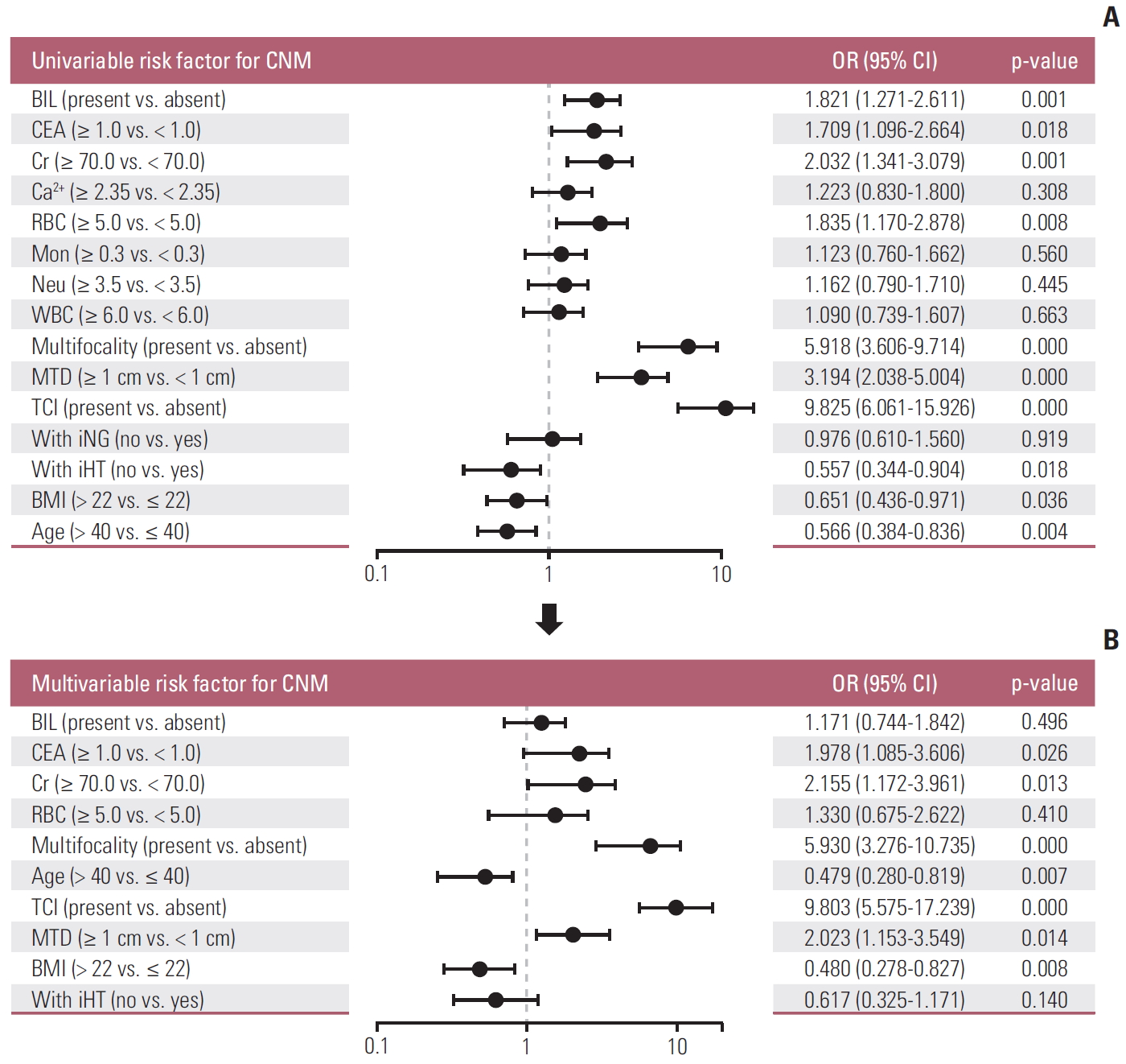
In the univariate analysis, bilaterality (p < 0.001), CEA (p=0.018), Cr (p < 0.001), red blood cell (p=0.008), multifocality (US) (p < 0.001), maximum tumor diameter (MTD, US) (p < 0.001), TCI (US) (p < 0.001), ipsilateral Hashimoto thyroiditis (p=0.018), BMI (p=0.018), and age (p=0.004) have association with CNM (Fig. 1A). Multivariate logistic regression modeling was further conducted to screen for significant variables associated with CNM. Results were as follows: CEA > 1 ng/mL, Cr > 70 μmol/L, multifocality, age < 40 years, TCI, MTD > 1 cm, BMI < 22 (Fig. 1B).
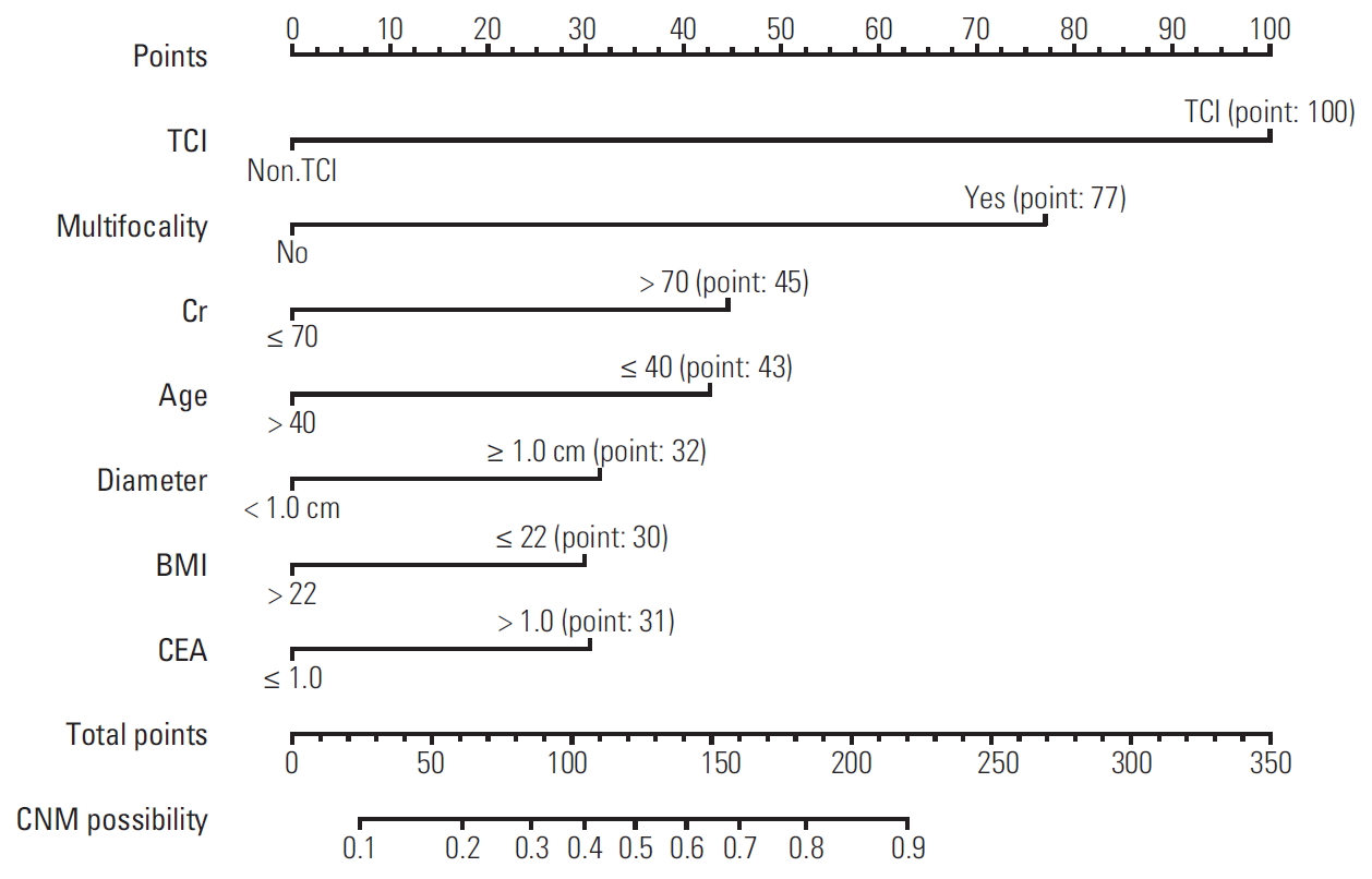
3. Nomogram for predicting likelihood of CNM in PTC patients
Based on the independent factors screened through multivariate analysis, a nomogram was established for predicting individual risk of CNM. The risk of each factor including CEA, Cr, multifocality, age, TCI, and BMI was quantified in our predicting model (score of each factor was shown in Fig. 2) to predict the presence of CNM in PTC patients.
4. Internal and external validation of the nomogram
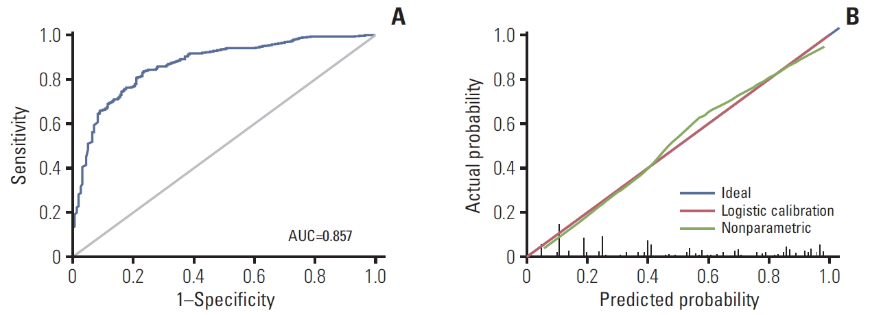
To evaluate our nomogram’s ability to predict CNM in PTC patients, we conducted an internal validation using 1,000 bootstrap resamples. A C-index of 0.857 (95% CI, 0.821 to 0.894) was achieved, and a similar C-index 0.854 (95% CI, 0.843 to 0.867) was acquired after 1,000 bootstrapping, confirming its satisfactory accuracy in predicting central lymph nodes involvement. ROC curve and AUC are presented in Fig. 3A. Furthermore, we also conducted a calibration plot for our newly-established model, and a favorable agreement was shown between the actual and estimated probability of CNM (Fig. 3B). The external validation population comprised of 186 patients (122 females) with a 53% overall rate of nodal metastases. The mean age was 41.8 years with a range of 19-72 years. When applied to the original model, the external validation data set produced a C-index of 0.825 (95% CI, 0.793 to 0.857) (Fig. 4).
5. Validation of the predictive effect of serum factors
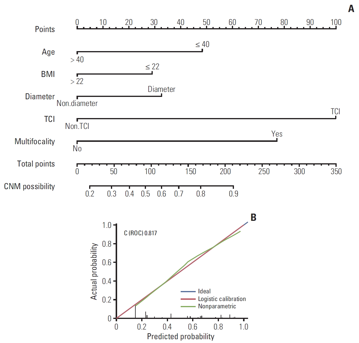
In order to evaluate the effect of serum indicators (CEA and Cr) we included in this prediction model, we excluded two serum indicators and reproduced a contrast nomogram (Fig. 5A), with a C-index of 0.817 (Fig. 5B), proving that these two factors we included were significant and could improve our nomogram’s ability to predict CNM.
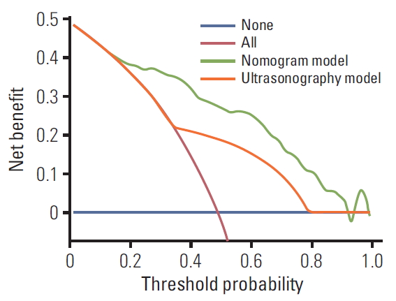
6. Comparing two models in predicting CNM based upon a decision curve analysis
A DCA was performed to compare the predictive ability between our nomogram and the US model. The US model used the data from the preoperative ultrasonographic diagnosis of CNM in our patient cohort (n=1,252). The DCA showed that the prediction ability of our nomogram is superior to US in detecting CNM for PTC patients (Fig. 6).
7. Novel risk stratification according to the scores obtained by the nomogram
Every variable included in our nomogram had its corresponding risk point and total risk scores calculated for all patients to quantitatively predict their individual risk of CNM. To stratify patients according to their risk scores, four cut-off values were selected. Five subgroups were thus established: (1) extreme low-risk group (patients with a CNM score of ≤ 50, n=137), (2) low-risk group (50 < risk score ≤ 100, n=311), (3) moderate-risk group (100 < risk score ≤ 150, n=305), (4) high-risk group (150 < risk score ≤ 200, n=184), and (5) extreme high-risk group (patients with a CNM score of > 200, n=315).
The rates of CNM for extreme low, low, moderate, high, extreme high-risk groups were 8.8%, 17.7%, 41.3%, 77.2%, and 89.8%, respectively. Differences were significant between these subgroups (p < 0.001) (Table 2).
Increasing incidence of thyroid cancer has been reported over recent years, and surgical resection is generally accepted to be the most effective treatment for PTC [11], yet the role of prophylactic CLND in PTC continues to be debated. Advocates point that prophylactic CLND in cN0 patient reduces local recurrence which contributes to less hazardous reoperative surgery [6,12,13]. In our country, given the combination of higher CNM risks in Asian patients and unreliability of preoperative examinations in detecting CNM [14], most institutions prefer thyroidectomy with prophylactic CLND. In this study, the rate of patients who received prophylactic CLND was 96.1% (1,663/1,731).
Although there is increasing support for prophylactic CLND given its benefits for recurrence and survival, many clinicians worldwide still opt for therapeutic CLND only, due to potential risks of prophylactic CLND. These could include permanent hypoparathyroidism, recurrent laryngeal nerve injury, and other postoperative complications [15-17]. Furthermore, in the American Thyroid Association guidelines (2015), “less is more” seemed to be the theme running throughout the consensus, recommending less extensive surgeries, less radioactive iodine and less surveillance testing [9,18]. In addition, the importance of regional lymph nodes for cancer survival is stressed in immunotherapy [19], as preservation of normal lymph nodes in meaningful for follow-up treatments. Thus, accurately assessing risks of CNM may play an important role in surgical options, for it is the deciding factor in whether prophylactic CLND should be performed.
Our study aimed to develop a nomogram to predict the likelihood of CNM in PTC patients. A CNM rate of 49.4% was identified, which is in accordance with figures of 40-58% reported by other similar works [20,21]. Among these patients, 158 patients (25.6%) were observed to have LLNM, while 43 patients (3.4%) had skip metastasis (i.e., had LLNM without CNM) which is similar to the observation made by Thompson et al. [6,22]. Given the low prevalence of skip metastasis, most LLNM will be accompanied by CNM, and a routine CLND will be performed; thus, we question the significance of including LLNM as a risk factor for CNM suggested by Wang et al. [23]. However, their results match our finding where there is a significant association between CNM and LLNM, and we will report this clinical research in our following work.
Referencing similar works on predicting CNM, our research confirms their results and takes it a step further. The nomogram developed by Thompson et al. [6] was based on 914 patients and had good discrimination with a C-index of 0.764, while only four variables were considered. The nomograms established by Wang et al. [23] and Kim et al. [24] enrolled larger patient cohorts, yet their discrimination presented a C-index of 0.711 to 0.721, which was weaker than this study. In addition, the equal split of the variables (age and tumor size) in Kim et al.’s work [24] is also quite puzzling given the nonlinear relationship between the variable and CNM according to Thompson’s article [6], which raised further questions about the representative of their nomogram for the general population.
There were a few references on the association between BMI and CNM [25,26], yet our results were quite different to previous findings. Wu et al. [25] found that increased BMI was associated with lymph node metastasis by analyzing a cohort of 796 PTC patients, while we observed that BMI less than 22 kg/m2 was the risk factor for CNM of PTC patients. This difference may be caused by disparate ways of BMI grouping or and the diagnose distinction of lymph node metastasis. However, we believe our nomogram showed a better predictive accuracy (AUC, 0.854) based on greater amount of samples of 1,252 PTC patients.
In this study, we took TCI diagnosed by preoperative US as variable instead of pathological TCI due to the lack of pathologically uniform standard (whether TCI or extrathyroidal extension [ETE]) in our institutions. This stems from a lack of definite definition for TCI or ETE in thyroid pathology [27]. Preoperative US TCI was identified in 39.5% cases, while other studies repor-ted that pathological TCI was present in around 30%-53% cases [6,28,29]. Interestingly, we observed TCI to be the most sensitive characteristic in our nomogram. Several works have proven the connections between TCI or ETE and CNM [23,24] and Thompson et al. [6] found wider spread ETE had a higher likelihood of lymph node metastasis compared to mere capsular invasion.
Multifocality and large tumor size have frequently been shown to promote CNM of PTC patients [2,6,12,20,23,24]. In our research, we obtained similar findings to previous studies, shown in our nomogram (Fig. 2), in which multifocality and tumor size larger than 1 cm both played an important role.
Preoperative serum index is available in most institutions, yet their association with CNM of PTC patients seems to be overlooked. We gathered all the pre-operation clinical serum index including blood routine examination, serum hepatorenal function, serum electrolyte, serum thyroid function, parathyroid hormone and CEA. Interestingly, we found serum Cr and CEA did contribute to our nomogram, suggesting PTC patients with Cr over 70 μmol/L or CEA over 1 ng/mL might suffer a higher risk of CNM.
Above all, this nomogram made it possible to score the likelihood of CNM in PTC patients before operation, which showed a significant advantage over preoperative US (Fig. 6). By gathering readily available clinical characteristics, we divided PTC patients into five quantified risk stratification in our nomogram (Table 2) so that it is easier to use clinically. Using this risk stratification table, clinicians may be better informed in their assessment of the risk factors.
There are several limitations of this study which we hope to address in our following research. Although we gathered patient cohort from two institutions, our sample was not as large as the two recent researches [23,24]. In addition, only 523 patients’ BRAF mutation reports were available and fewer patients received a postoperative immunohistochemical examinations. Future predictive models may incorporate microRNA and PTC subtypes, as well as immunohistochemical biopsy and genotype of FNA samples, which are not available at present. Furthermore, we were able to incorporate several new variables that are significant for prediction of CNM. This implies that there may be potential variables waiting to be discovered that could make the nomogram more complete.
Using available pre-operative variables, we were able to construct a nomogram that stratify PTC patients into five groups that possess different CNM risk levels. This provides guidance for whether a patient should receive CLND on a case by case basis, and promotes balanced approach between avoiding CLND complications and maximizing survival/lowering recurrence rates.

Conflicts of interest relevant to this article was not reported.

Acknowledgements
This research was supported by the Shanghai Municipal Science and Technology Commission(19441905400); Shanghai Jiaotong University(YG2019ZDA15).
Fig. 1.
Univariate (A) and multivariate (B) logistic regression of factors associated with central lymph node metastasis (CNM). BIL, bilirubin; CEA, carcinoembryonic antigen; Cr, creatinine; RBC, red blood cell; Mon, monocyte; Neu, neutrophil; WBC, white blood cell; MTD, maximum tumor diameter; TCI, thyroid capsular invasion; iNG, ipsilateral Nodular Goiter; iHT, ipsilateral Hashimoto thyroiditis; BMI, body mass index.
crt-2020-254f1.jpg
Fig. 2.
The nomogram for predicting risk of possible central lymph node metastasis (CNM) in papillary thyroid carcinoma patients. TCI, thyroid capsular invasion; Cr, creatinine; BMI, body mass index; CEA, carcinoembryonic antigen.
crt-2020-254f2.jpg
Fig. 3.
Evaluation and Validation of the nomogram. (A) The receiver operating characteristics (ROC) curve and area under the ROC curve (AUC) of the nomogram. (B) The calibration curve of the nomogram for predicting possible central lymph node metastasis. Actual probability is plotted on the y-axis, and nomogram predicted probability on the x-axis.
crt-2020-254f3.jpg
Fig. 4.
The calibration curve of the nomogram for external validation set. ROC, receiver operating characteristics.
crt-2020-254f4.jpg
Fig. 5.
(A) Nomogram without serum index for predicting central lymph node metastasis (CNM) in papillary thyroid carcinoma (PTC) patients. (B) The calibration curve of the nomogram excluding serum index. BMI, body mass index; TCI, thyroid capsular invasion; ROC, receiver operating characteristics.
crt-2020-254f5.jpg
Fig. 6.
Decision curve analysis for comparing our nomogram and ultrasonography in predicting central lymph node metastasis in papillary thyroid carcinoma patients.
crt-2020-254f6.jpg
Table 1.
Demographics and clinical characteristics of the cohort
Variable Value
Age (yr)
 Mean 43.1
BMI (kg/m2)
 Mean 23.81
Sex
 Male 447 (35.7)
 Female 805 (64.3)
Size of largest lesion (US)
 Mean 0.86
 Medium 0.7
Thyroid capsule invasion (US) 495 (39.5)
Multifocality (US) 373 (29.8)
Bilateral disease 243 (19.4)
Serum index (mean)
 CEA 1.42
 Creatinine 64
CNM 618 (49.4)
Central lymph node
 Mean harvested 7.1
 Mean positive 4.3
LLNM 158 (12.6)

Vaues are presented as number (%) unless otherwise indicated. BMI, body mass index. US, ultrasonography; CEA, carcinoembryonic antigen; CNM, central lymph node metastasis; LLNM, lateral lymph node metastasis.

Table 2.
CNM risk stratification of PTC patients based on the model database
Nomogram ELR
LR
MR
HR
HER
Total
0-50 50-100 100-150 150-200 > 200
No CNM Value 125 256 179 42 32 634
% 91.2 82.3 58.7 22.8 10.2 50.6
CNM Value 12 55 126 142 283 618
% 8.8 17.7 41.3 77.2 89.8 49.4
Total Value 137 311 305 184 315 1,252
% 100 100 100 100 100 100

CNM, central lymph node metastasis; PTC, papillary thyroid carcinoma; ELR, extreme low risk; LR, low risk; MR, moderated risk; HR, high risk; HER, extreme high risk.

  • 1. Maino F, Forleo R, Pacini F. Prognostic indicators for papillary thyroid carcinoma. Expert Rev Endocrinol Metab. 2017;12:101–8. ArticlePubMed
  • 2. Al Afif A, Williams BA, Rigby MH, Bullock MJ, Taylor SM, Trites J, et al. Multifocal papillary thyroid cancer increases the risk of central lymph node metastasis. Thyroid. 2015;25:1008–12. ArticlePubMed
  • 3. Bernet V. Approach to the patient with incidental papillary microcarcinoma. J Clin Endocrinol Metab. 2010;95:3586–92. ArticlePubMedPDF
  • 4. Stulak JM, Grant CS, Farley DR, Thompson GB, van Heerden JA, Hay ID, et al. Value of preoperative ultrasonography in the surgical management of initial and reoperative papillary thyroid cancer. Arch Surg. 2006;141:489–94. ArticlePubMed
  • 5. Kim SK, Woo JW, Lee JH, Park I, Choe JH, Kim JH, et al. Prophylactic central neck dissection might not be necessary in papillary thyroid carcinoma: analysis of 11,569 cases from a single institution. J Am Coll Surg. 2016;222:853–64. ArticlePubMed
  • 6. Thompson AM, Turner RM, Hayen A, Aniss A, Jalaty S, Learoyd DL, et al. A preoperative nomogram for the prediction of ipsilateral central compartment lymph node metastases in papillary thyroid cancer. Thyroid. 2014;24:675–82. ArticlePubMedPMC
  • 7. Egelmeer AG, Velazquez ER, de Jong JM, Oberije C, Geussens Y, Nuyts S, et al. Development and validation of a nomogram for prediction of survival and local control in laryngeal carcinoma patients treated with radiotherapy alone: a cohort study based on 994 patients. Radiother Oncol. 2011;100:108–15. ArticlePubMed
  • 8. Wan G, Gao F, Chen J, Li Y, Geng M, Sun L, et al. Nomogram prediction of individual prognosis of patients with hepatocellular carcinoma. BMC Cancer. 2017;17:91.ArticlePubMedPMCPDF
  • 9. Tuttle RM, Haugen B, Perrier ND. Updated American Joint Committee on Cancer/tumor-node-metastasis staging system for differentiated and anaplastic thyroid cancer (eighth edition): what changed and why? Thyroid. 2017;27:751–6. ArticlePubMedPMC
  • 10. Mo S, Dai W, Xiang W, Li Q, Wang R, Cai G. Predictive factors of synchronous colorectal peritoneal metastases: Development of a nomogram and study of its utilities using decision curve analysis. Int J Surg. 2018;54(Pt A):149–55. ArticlePubMed
  • 11. Pellegriti G, Frasca F, Regalbuto C, Squatrito S, Vigneri R. Worldwide increasing incidence of thyroid cancer: update on epidemiology and risk factors. J Cancer Epidemiol. 2013;2013:965212.ArticlePubMedPMCPDF
  • 12. Perrino M, Vannucchi G, Vicentini L, Cantoni G, Dazzi D, Colombo C, et al. Outcome predictors and impact of central node dissection and radiometabolic treatments in papillary thyroid cancers < or =2 cm. Endocr Relat Cancer. 2009;16:201–10. ArticlePubMed
  • 13. Popadich A, Levin O, Lee JC, Smooke-Praw S, Ro K, Fazel M, et al. A multicenter cohort study of total thyroidectomy and routine central lymph node dissection for cN0 papillary thyroid cancer. Surgery. 2011;150:1048–57. ArticlePubMed
  • 14. Goropoulos A, Karamoshos K, Christodoulou A, Ntitsias T, Paulou K, Samaras A, et al. Value of the cervical compartments in the surgical treatment of papillary thyroid carcinoma. World J Surg. 2004;28:1275–81. ArticlePubMedPDF
  • 15. Lee YS, Kim SW, Kim SW, Kim SK, Kang HS, Lee ES, et al. Extent of routine central lymph node dissection with small papillary thyroid carcinoma. World J Surg. 2007;31:1954–9. ArticlePubMedPDF
  • 16. Moo TA, McGill J, Allendorf J, Lee J, Fahey T 3rd, Zarnegar R. Impact of prophylactic central neck lymph node dissection on early recurrence in papillary thyroid carcinoma. World J Surg. 2010;34:1187–91. ArticlePubMedPDF
  • 17. Sancho JJ, Lennard TW, Paunovic I, Triponez F, Sitges-Serra A. Prophylactic central neck disection in papillary thyroid cancer: a consensus report of the European Society of Endocrine Surgeons (ESES). Langenbecks Arch Surg. 2014;399:155–63. ArticlePubMedPDF
  • 18. Kim BW, Yousman W, Wong WX, Cheng C, McAninch EA. Less is more: comparing the 2015 and 2009 American Thyroid Association guidelines for thyroid nodules and cancer. Thyroid. 2016;26:759–64. ArticlePubMed
  • 19. Thomas SN, Rohner NA, Edwards EE. Implications of lymphatic transport to lymph nodes in immunity and immunotherapy. Annu Rev Biomed Eng. 2016;18:207–33. ArticlePubMedPMC
  • 20. Lee YS, Lim YS, Woo CG, Lee JC, Wang SG, Lee KD, et al. PP049: growth pattern of micropapillary thyroid cancer without extrathyroidal extension predicts central lymph node metastasis. Oral Oncol. 2013;49(Suppl 1):S110.Article
  • 21. Usluogullari CA, Onal ED, Ozdemir E, Ucler R, Kiyak G, Ersoy PE, et al. A retrospective analysis of prognostic factors predictive of lymph-node metastasis and recurrence in thyroid papillary microcarcinoma. Minerva Endocrinol. 2015;40:15–22. PubMed
  • 22. Grebe SK, Hay ID. Thyroid cancer nodal metastases: biologic significance and therapeutic considerations. Surg Oncol Clin N Am. 1996;5:43–63. ArticlePubMed
  • 23. Wang Y, Guan Q, Xiang J. Nomogram for predicting central lymph node metastasis in papillary thyroid microcarcinoma: a retrospective cohort study of 8668 patients. Int J Surg. 2018;55:98–102. ArticlePubMed
  • 24. Kim SK, Chai YJ, Park I, Woo JW, Lee JH, Lee KE, et al. Nomogram for predicting central node metastasis in papillary thyroid carcinoma. J Surg Oncol. 2017;115:266–72. ArticlePubMed
  • 25. Wu C, Wang L, Chen W, Zou S, Yang A. Associations between body mass index and lymph node metastases of patients with papillary thyroid cancer: a retrospective study. Medicine (Baltimore). 2017;96:e6202ArticlePubMedPMC
  • 26. Dieringer P, Klass EM, Caine B, Smith-Gagen J. Associations between body mass and papillary thyroid cancer stage and tumor size: a population-based study. J Cancer Res Clin Oncol. 2015;141:93–8. ArticlePubMedPDF
  • 27. Mete O, Rotstein L, Asa SL. Controversies in thyroid pathology: thyroid capsule invasion and extrathyroidal extension. Ann Surg Oncol. 2010;17:386–91. ArticlePubMedPDF
  • 28. Kim ES, Lim DJ, Lee K, Jung CK, Bae JS, Jung SL, et al. Absence of galectin-3 immunostaining in fine-needle aspiration cytology specimens from papillary thyroid carcinoma is associated with favorable pathological indices. Thyroid. 2012;22:1244–50. ArticlePubMed
  • 29. Furlan JC, Bedard YC, Rosen IB. Significance of tumor capsular invasion in well-differentiated thyroid carcinomas. Am Surg. 2007;73:484–91. ArticlePubMed

Figure & Data

REFERENCES

    Citations

    Citations to this article as recorded by  
    • Risk nomogram for papillary thyroid microcarcinoma with central lymph node metastasis and postoperative thyroid function follow-up
      Yuting Huang, Pengwei Lou, Hui Li, Yinhui Li, Li Ma, Kai Wang
      Frontiers in Endocrinology.2024;[Epub]     CrossRef
    • Ultrasonic Feature Prediction of Large-Number Central Lymph Node Metastasis in Clinically Node-Negative Solitary Papillary Thyroid Carcinoma
      Weihan Xiao, Xiaomin Hu, Chaoxue Zhang, Xiachuan Qin
      Endocrine Research.2023; 48(4): 112.     CrossRef
    • Nomogram for predicting central lymph node metastasis in T1-T2 papillary thyroid cancer with no lateral lymph node metastasis
      Yubo Sun, Wei Sun, Jingzhe Xiang, Hao Zhang
      Frontiers in Endocrinology.2023;[Epub]     CrossRef
    • An integrated nomogram combining deep learning, clinical characteristics and ultrasound features for predicting central lymph node metastasis in papillary thyroid cancer: A multicenter study
      Luchen Chang, Yanqiu Zhang, Jialin Zhu, Linfei Hu, Xiaoqing Wang, Haozhi Zhang, Qing Gu, Xiaoyu Chen, Sheng Zhang, Ming Gao, Xi Wei
      Frontiers in Endocrinology.2023;[Epub]     CrossRef
    • Central and lateral neck involvement in papillary thyroid carcinoma patients with or without thyroid capsular invasion: A multi-center analysis
      Zheyu Yang, Yu Heng, Jian Zhou, Lei Tao, Wei Cai
      Frontiers in Endocrinology.2023;[Epub]     CrossRef
    • Development and validation of nomograms for predicting the risk of central lymph node metastasis of solitary papillary thyroid carcinoma of the isthmus
      Yonghao Li, Xuefei Gao, Tiantian Guo, Jing Liu
      Journal of Cancer Research and Clinical Oncology.2023; 149(16): 14853.     CrossRef
    • Development and validation of an individualized nomogram for predicting the high-volume (> 5) central lymph node metastasis in papillary thyroid microcarcinoma
      X. Wei, Y. Min, Y. Feng, D. He, X. Zeng, Y. Huang, S. Fan, H. Chen, J. Chen, K. Xiang, H. Luo, G. Yin, D. Hu
      Journal of Endocrinological Investigation.2022; 45(3): 507.     CrossRef
    • Preoperative Prediction of Central Cervical Lymph Node Metastasis in Fine-Needle Aspiration Reporting Suspicious Papillary Thyroid Cancer or Papillary Thyroid Cancer Without Lateral Neck Metastasis
      Kai Zhang, Lang Qian, Jieying Chen, Qian Zhu, Cai Chang
      Frontiers in Oncology.2022;[Epub]     CrossRef
    • Relationship between subgroups of central and lateral lymph node metastasis in clinically node-negative papillary thyroid carcinoma
      Jing Zhou, Da-Xue Li, Han Gao, Xin-Liang Su
      World Journal of Clinical Cases.2022; 10(12): 3709.     CrossRef
    • Hashimoto’s Thyroiditis Is Associated With Central Lymph Node Metastasis in Classical Papillary Thyroid Cancer: Analysis from a High-Volume Single-Center Experience
      Bin Zeng, Yu Min, Yang Feng, Ke Xiang, Hang Chen, Zijing Lin
      Frontiers in Endocrinology.2022;[Epub]     CrossRef
    • Nomogram model based on preoperative serum thyroglobulin and clinical characteristics of papillary thyroid carcinoma to predict cervical lymph node metastasis
      Qungang Chang, Jieming Zhang, Yaqian Wang, Hongqiang Li, Xin Du, Daohong Zuo, Detao Yin
      Frontiers in Endocrinology.2022;[Epub]     CrossRef
    • Ultrasound-based radiomics nomogram combined with clinical features for the prediction of central lymph node metastasis in papillary thyroid carcinoma patients with Hashimoto’s thyroiditis
      Peile Jin, Jifan Chen, Yiping Dong, Chengyue Zhang, Yajun Chen, Cong Zhang, Fuqiang Qiu, Chao Zhang, Pintong Huang
      Frontiers in Endocrinology.2022;[Epub]     CrossRef
    • Lateral Involvement in Different Sized Papillary Thyroid Carcinomas Patients with Central Lymph Node Metastasis: A Multi-Center Analysis
      Yu Heng, Zheyu Yang, Pengyu Cao, Xi Cheng, Lei Tao
      Journal of Clinical Medicine.2022; 11(17): 4975.     CrossRef
    • Risk factors and prediction models of lymph node metastasis in papillary thyroid carcinoma based on clinical and imaging characteristics
      Yanyuan Deng, Jie Zhang, Jiao Wang, Jinying Wang, Junping Zhang, Lulu Guan, Shasha He, Xiudan Han, Wei Cai, Jixiong Xu
      Postgraduate Medicine.2022;[Epub]     CrossRef
    • A prognostic nomogram for papillary thyroid cancer lymph node metastasis based on immune score
      Yihua Lu, Kai Qian, Mengjia Fei, Kai Guo, Yuan Shi, Zhuoying Wang
      Frontiers in Endocrinology.2022;[Epub]     CrossRef
    • Risk factors for central and lateral lymph node metastasis in papillary thyroid carcinoma
      Ji Hyun Ahn, Hee Kyung Chang
      Kosin Medical Journal.2022; 37(4): 311.     CrossRef
    • Construction and validation of a nomogram for predicting cervical lymph node metastasis in classic papillary thyroid carcinoma
      Y. Feng, Y. Min, H. Chen, K. Xiang, X. Wang, G. Yin
      Journal of Endocrinological Investigation.2021; 44(10): 2203.     CrossRef
    • Application of Machine Learning Algorithms to Predict Central Lymph Node Metastasis in T1-T2, Non-invasive, and Clinically Node Negative Papillary Thyroid Carcinoma
      Jiang Zhu, Jinxin Zheng, Longfei Li, Rui Huang, Haoyu Ren, Denghui Wang, Zhijun Dai, Xinliang Su
      Frontiers in Medicine.2021;[Epub]     CrossRef
    • Preoperative Predictors of Lymph Node Metastasis in Colon Cancer
      Yansong Xu, Yi Chen, Chenyan Long, Huage Zhong, Fangfang Liang, Ling-xu Huang, Chuanyi Wei, Shaolong Lu, Weizhong Tang
      Frontiers in Oncology.2021;[Epub]     CrossRef
    • Preoperatively Predicting the Central Lymph Node Metastasis for Papillary Thyroid Cancer Patients With Hashimoto’s Thyroiditis
      Yu Min, Yizhou Huang, Minjie Wei, Xiaoyuan Wei, Hang Chen, Xing Wang, Jialin Chen, Ke Xiang, Yang Feng, Guobing Yin
      Frontiers in Endocrinology.2021;[Epub]     CrossRef
    • Reevaluation of Criteria and Establishment of Models for Total Thyroidectomy in Differentiated Thyroid Cancer
      Zhenghao Wu, Yunxiao Xiao, Jie Ming, Yiquan Xiong, Shuntao Wang, Shengnan Ruan, Tao Huang
      Frontiers in Oncology.2021;[Epub]     CrossRef
    • Influencing Factors and Cumulative Risk Analysis of Cervical Lymph Node Metastasis of Papillary Thyroid Microcarcinoma
      Yirong Yin, Xiang Xu, Liyan Shen, Wenjuan Zhao, Hongcui Diao, Chengqian Li
      Frontiers in Oncology.2021;[Epub]     CrossRef
    • A Specific Predicting Model for Screening Skip Metastasis From Patients With Negative Central Lymph Nodes Metastasis in Papillary Thyroid Cancer
      Zheyu Yang, Yu Heng, Qiwu Zhao, Zichao Cao, Lei Tao, Weihua Qiu, Wei Cai
      Frontiers in Endocrinology.2021;[Epub]     CrossRef
    • Computed Tomography-Based Radiomics Model to Predict Central Cervical Lymph Node Metastases in Papillary Thyroid Carcinoma: A Multicenter Study
      Jingjing Li, Xinxin Wu, Ning Mao, Guibin Zheng, Haicheng Zhang, Yakui Mou, Chuanliang Jia, Jia Mi, Xicheng Song
      Frontiers in Endocrinology.2021;[Epub]     CrossRef
    • Nomogram Including Elastography for Prediction of Contralateral Central Lymph Node Metastasis in Solitary Papillary Thyroid Carcinoma Preoperatively


      Ning Li, Ju-hua He, Chao Song, Li-chun Yang, Hong-jiang Zhang, Zhi-hai Li
      Cancer Management and Research.2020; Volume 12: 10789.     CrossRef

    • PubReader PubReader
    • ePub LinkePub Link
    • Cite
      CITE
      export Copy Download
      Close
      Download Citation
      Download a citation file in RIS format that can be imported by all major citation management software, including EndNote, ProCite, RefWorks, and Reference Manager.

      Format:
      • RIS — For EndNote, ProCite, RefWorks, and most other reference management software
      • BibTeX — For JabRef, BibDesk, and other BibTeX-specific software
      Include:
      • Citation for the content below
      Nomogram for Predicting Central Lymph Node Metastasis in Papillary Thyroid Cancer: A Retrospective Cohort Study of Two Clinical Centers
      Cancer Res Treat. 2020;52(4):1010-1018.   Published online June 9, 2020
      Close
    • XML DownloadXML Download
    Nomogram for Predicting Central Lymph Node Metastasis in Papillary Thyroid Cancer: A Retrospective Cohort Study of Two Clinical Centers
    Image Image Image Image Image Image
    Fig. 1. Univariate (A) and multivariate (B) logistic regression of factors associated with central lymph node metastasis (CNM). BIL, bilirubin; CEA, carcinoembryonic antigen; Cr, creatinine; RBC, red blood cell; Mon, monocyte; Neu, neutrophil; WBC, white blood cell; MTD, maximum tumor diameter; TCI, thyroid capsular invasion; iNG, ipsilateral Nodular Goiter; iHT, ipsilateral Hashimoto thyroiditis; BMI, body mass index.
    Fig. 2. The nomogram for predicting risk of possible central lymph node metastasis (CNM) in papillary thyroid carcinoma patients. TCI, thyroid capsular invasion; Cr, creatinine; BMI, body mass index; CEA, carcinoembryonic antigen.
    Fig. 3. Evaluation and Validation of the nomogram. (A) The receiver operating characteristics (ROC) curve and area under the ROC curve (AUC) of the nomogram. (B) The calibration curve of the nomogram for predicting possible central lymph node metastasis. Actual probability is plotted on the y-axis, and nomogram predicted probability on the x-axis.
    Fig. 4. The calibration curve of the nomogram for external validation set. ROC, receiver operating characteristics.
    Fig. 5. (A) Nomogram without serum index for predicting central lymph node metastasis (CNM) in papillary thyroid carcinoma (PTC) patients. (B) The calibration curve of the nomogram excluding serum index. BMI, body mass index; TCI, thyroid capsular invasion; ROC, receiver operating characteristics.
    Fig. 6. Decision curve analysis for comparing our nomogram and ultrasonography in predicting central lymph node metastasis in papillary thyroid carcinoma patients.
    Nomogram for Predicting Central Lymph Node Metastasis in Papillary Thyroid Cancer: A Retrospective Cohort Study of Two Clinical Centers
    Variable Value
    Age (yr)
     Mean 43.1
    BMI (kg/m2)
     Mean 23.81
    Sex
     Male 447 (35.7)
     Female 805 (64.3)
    Size of largest lesion (US)
     Mean 0.86
     Medium 0.7
    Thyroid capsule invasion (US) 495 (39.5)
    Multifocality (US) 373 (29.8)
    Bilateral disease 243 (19.4)
    Serum index (mean)
     CEA 1.42
     Creatinine 64
    CNM 618 (49.4)
    Central lymph node
     Mean harvested 7.1
     Mean positive 4.3
    LLNM 158 (12.6)
    Nomogram ELR
    LR
    MR
    HR
    HER
    Total
    0-50 50-100 100-150 150-200 > 200
    No CNM Value 125 256 179 42 32 634
    % 91.2 82.3 58.7 22.8 10.2 50.6
    CNM Value 12 55 126 142 283 618
    % 8.8 17.7 41.3 77.2 89.8 49.4
    Total Value 137 311 305 184 315 1,252
    % 100 100 100 100 100 100
    Table 1. Demographics and clinical characteristics of the cohort

    Vaues are presented as number (%) unless otherwise indicated. BMI, body mass index. US, ultrasonography; CEA, carcinoembryonic antigen; CNM, central lymph node metastasis; LLNM, lateral lymph node metastasis.

    Table 2. CNM risk stratification of PTC patients based on the model database

    CNM, central lymph node metastasis; PTC, papillary thyroid carcinoma; ELR, extreme low risk; LR, low risk; MR, moderated risk; HR, high risk; HER, extreme high risk.


    Cancer Res Treat : Cancer Research and Treatment
    Close layer
    TOP