Skip Navigation
Skip to contents

Cancer Res Treat : Cancer Research and Treatment

OPEN ACCESS

Articles

Page Path
HOME > Cancer Res Treat > Volume 48(2); 2016 > Article
Original Article Radiation-Induced Sarcoma: A 15-Year Experience in a Single Large Tertiary Referral Center
Kyung Su Kim, MD1, Ji Hyun Chang, MD2, Noorie Choi, MD1, Han-Soo Kim, MD, PhD3, Ilkyu Han, MD, PhD3, Kyung Chul Moon, MD, PhD4, Il Han Kim, MD, PhD1, Hak Jae Kim, MD, PhD1,
Cancer Research and Treatment : Official Journal of Korean Cancer Association 2016;48(2):650-657.
DOI: https://doi.org/10.4143/crt.2015.171
Published online: September 9, 2015

1Department of Radiation Oncology, Seoul National University College of Medicine, Seoul, Korea

2Department of Radiation Oncology, Seoul St. Mary's Hospital, College of Medicine, The Catholic University of Korea, Seoul, Korea

3Department of Orthopedic Surgery, Seoul National University College of Medicine, Seoul, Korea

4Department of Pathology, Seoul National University College of Medicine, Seoul, Korea

Correspondence: Hak Jae Kim, MD, PhD  Department of Radiation Oncology, Seoul National University College of Medicine, 103 Daehak-ro, Jongno-gu, Seoul 03080, Korea 
Tel: 82-2-2072-2520 Fax: 82-2-765-3317 E-mail: khjae@snu.ac.kr
*Kyung Su Kim and Ji Hyun Chang contributed equally to this work.
• Received: May 19, 2015   • Accepted: August 17, 2015

Copyright © 2016 by the Korean Cancer Association

This is an Open Access article distributed under the terms of the Creative Commons Attribution Non-Commercial License (http://creativecommons.org/licenses/by-nc/3.0/) which permits unrestricted non-commercial use, distribution, and reproduction in any medium, provided the original work is properly cited.

  • 14,048 Views
  • 197 Download
  • 29 Web of Science
  • 36 Crossref
  • 31 Scopus
prev next
  • Purpose
    The purpose of this study is to report on the incidence and the experience in management of radiation-induced sarcoma (RIS) at a large single center in Korea for 15 years.
  • Materials and Methods
    We retrospectively reviewed the sarcoma registry of a large institution from January 2000 to April 2014.
  • Results
    Out of the 3,674 patients listed in the registry, 33 patients (0.9%) diagnosed with RIS were identified. The median latency of RIS was 12.1 years. The number of cases of RIS increased from four cases in the years 2000-2003 to 14 cases in the years 2012-2014. The most common histology was osteosarcoma (36.4%). The median follow-up period was 23.1 months, the median overall survival (OS) of all patients was 2.9 years, and their 5-year survival rate was 44.7%. Univariate and multivariate analyses showed association of the age at diagnosis (p=0.01) and the treatment aim (p=0.001) with the OS. The median OS and the 5-year survival rate of patients treated with curative surgery (n=19) were 9.6 years and 65%, respectively, and of the conservatively treated patients, 0.7 years and 0% (n=14). Re-irradiation was delivered to nine patients, and radiation toxicity was observed in five patients.
  • Conclusion
    In this study, RIS accounted for 0.9% of the cases of sarcoma, with increasing incidence. Despite the association of curative resection with increased survival, it could be applied to only 58% of the patients. Considering the limited treatment options for RIS, conduct of a genetic study to identify the underlying mechanism of RIS is needed.
Radiation-induced sarcoma (RIS) is a rare late event after radiation therapy (RT), with a reported incidence of 0.03%- 0.2% in 5 years and overall up to 0.8% [1,2]. It is known to have a worse prognosis than sporadic soft tissue sarcoma (STS), with a 5-year disease-specific survival (DSS) rate of 32%-58% in recent reports [3-5]. Gladdy et al. [3] reported that RIS had a worse DSS than sporadic STS with a hazard ratio of 1.7. Dineen et al. [6] also recently reported a lower DSS of RIS than of sporadic STS.
Because RIS arises in the previously treated area, there are many limitations in its management. The major limitation is that RIS has lower resectability than sporadic sarcomas. Thijssens et al. [7] emphasized the low possibility of curative resection with data showing 62% R0 resection. In the study by Neuhaus et al. [8], R0-1 resection was achieved in approximately 37% of the patients and was the only factor showing significant correlation with the DSS. In addition, adjuvant RT for RIS should be administered with caution because of the higher risk of RT-related toxicities.
RT plays a critical role in the treatment of most cancers. Therefore, concerns about RIS are emerging among cancer survivors because the survival rate and the survival time after cancer treatment are increasing as a result of advanced treatments including the use of targeted agents.
Most studies of RIS in Asia are case reports, and significantly under-reported compared to western countries. RIS patients tend to be referred to a large tertiary center because of the surgical complexity, therefore we report herein the incidence and experience in management of RIS at a single large tertiary referral center for 15 years.
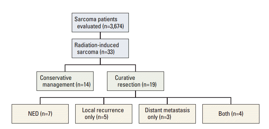
After receiving institutional review board approval, we retrospectively reviewed our institutional sarcoma registry from January 2000 to April 2014. Out of the 3,674 patients in the registry, we identified 33 patients who met the following criteria for RIS, which were modifications of Cahan’s criteria: (1) pathologic confirmation of sarcoma that was histologically unique from the primary cancer, (2) occurrence of the sarcoma in the field of radiation, and (3) radiation exposure at least 6 months before development of the sarcoma [9].
Medical records were reviewed for the patients’ age, sex, primary cancer treatment, RIS characteristics, RIS treatment, and follow-up data. The radiographic image of RIS and the histopathology report were assessed for identification of the tumor location, size, and histology. All available medical records from the follow-up were reviewed for evaluation of the disease status and toxicity. The toxicities from re-irradiation were evaluated using the Common Terminology Criteria for Adverse Events (CTCAE) ver. 4.0.
1. Statistical analysis
The overall survival rate was calculated from the time of the RIS diagnosis until death. Actuarial survival curves were plotted from the RIS diagnosis using the Kaplan-Meier method. A p-value less than 0.05 was considered to indicate a statistically significant difference. Analyses were performed using PASW ver. 18.0 (SPSS Inc., Chicago, IL).
1. Patients and tumor characteristics
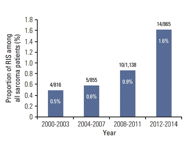
A total of 33 patients diagnosed with RIS were identified. The number of cases increased from four cases in the years 2000-2003 to 14 cases in the years 2012-2014 with corresponding proportion of RIS among sarcoma patients of 0.5% to 1.6% (Fig. 1).
A list of the characteristics of the 33 patients is shown in Table 1. Females accounted for 75.8% of the patients. The median age at the primary radiotherapy was 44 years (range, 0 to 71 years). Three patients were treated before age 20 (for bilateral retinoblastoma, Wilms’ tumor, and nasopharyngeal cancer), and 20 patients were treated in their 40s and 50s. The median age at the RIS diagnosis was 55 years (range, 22 to 85 years), and the median latency was 12.1 years (range, 1.9 to 28.8 years).
The most common type of primary cancer was breast cancer (n=9), followed by uterine cervical cancer (n=8), and head and neck cancer (n=7). The median prescribed dose for primary RT was 50.4 Gy. All patients received a dose greater than 45 Gy, with the exception of three patients with Wilms’ tumor, retinoblastoma, and lymphoma treated with 10.8 Gy, 36 Gy, and 30 Gy, respectively. Four patients underwent intracavitary brachytherapy in addition to external-beam RT for treatment of cervical cancer; 58% of patients underwent chemotherapy as their primary cancer treatment.
RIS was located in the trunk in 75.8% of the cases, and in the head and neck in 24.2%. The most common histology was osteosarcoma (36.4%), followed by malignant fibrous histiocytoma (33.3%) and angiosarcoma (6.1%). The median tumor size was 4.8 cm (range, 1.5 to 12.5 cm), and tumors were located deep in the muscle fascia in 27 cases (81.8%).
2. Treatment of RIS and outcome
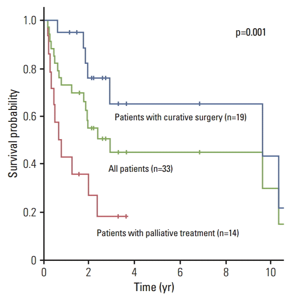
The median follow-up period was 23.1 months (range, 2.2 to 174.8 months). Nineteen patients underwent surgery with curative intent, while 14 patients were treated conservatively. A list of the details of the treatment of 19 patients with curative surgery is shown in Table 2. R0 resection was achieved, except in one patient. Four patients underwent neo-adjuvant chemotherapy, and six patients, adjuvant chemotherapy. One patient diagnosed with malignant histiocytoma in the mandible underwent adjuvant RT. The median overall survival period of all patients was 2.9 years, and their 5-year survival rate was 44.7%. In univariate and multivariate analyses, the age at diagnosis and the treatment aim showed association with the overall survival (Table 3). The median duration of the overall survival period and the 5-year survival rate of patients treated with curative surgery were 9.6 years and 65%, respectively, and that of the conservatively treated patients, 0.7 years and 0% (p=0.001) (Fig. 2).
The treatment outcomes are summarized in Fig. 3. Out of the 19 patients treated with curative resection, local recurrence occurred in nine patients (47%), and distant metastasis, in seven patients (37%).
3. Re-irradiation and toxicity
Nine patients underwent re-irradiation for various intents. Four patients were misdiagnosed with bone metastasis from their primary tumor and underwent palliative RT before biopsy. Three patients underwent palliative radiotherapy, one patient was treated with gamma knife surgery of the recurred mass, and one patient underwent adjuvant RT after curative surgery. Five patients, including the familial retinoblastoma patient who developed grade 4 skin necrosis after re-irradiation, showed radiation toxicity after re-irradiation (Table 4).
To the best of our knowledge, this is the first study reporting on the incidence and survival of RIS in Asia in a significant number of patients. Our data showed that, although rare, the number of patients diagnosed with RIS in our institution is consistently increasing with time. This may be due to the significant increase of RT usage in cancer therapy in South Korea since the first Cobalt-60 teletherapy installation in 1963. Patients undergoing RT increased from 22,000 in 1999 to 52,000 in 2011 [10]. The increased survival time of cancer patients could also be attributed to the increasing incidence of RIS.
RIS is known to have a worse prognosis than sporadic STS. Bjerkehagen et al. [5] investigated the question of whether or not a previous RT history is a worse prognosticator. In their analysis, a previous RT history was not a prognostic factor, but unlike sporadic STS, RIS tended to occur in a central location and to be associated with incomplete surgery, which are known poor prognosticators in sarcoma. As Cha et al. [11] stated, previous RT might obscure the anatomic and tumor planes, which might make radical resection challenging. Consistently in our data, only 57.6% of the patients could be treated with curative intent, and the survival outcome differed significantly according to the treatment aim.
In addition, the adjuvant RT for RIS is limited by the concern for possible complications regarding re-irradiation, which can hinder optimal RIS management. An adjuvant RT of 42-63 Gy is known to reduce local recurrence of STS after conservative surgery [12,13]. However, as shown in this study, RT for RIS results in a high rate of RT-related complications, which is inevitable in high-dose regions. Due to the rarity and the histologic diversity within RIS, it is difficult to analyze the efficacy of secondary adjuvant RT after curative resection of RIS. Extrapolating from sporadic STS, adjuvant RT should also be performed for RIS, but care is required in the process of consultation with the patient, RT planning, and prescription of the RT dose. Also, high-precision RT techniques with increased conformality, such as intensity-modulated radiation therapy (IMRT), should be used in such re-irradiation cases to minimize the toxicity.
The question of whether or not the use of IMRT, stereotactic body radiation therapy, or proton therapy would reduce RIS incidence is yet to be determined. From the follow-up observation data on atomic bomb survivors who showed no excess of sarcoma after exposure to radiation of a few Gy, we acknowledge that RIS is less likely to arise in very low-dose regions, and more likely to arise in intermediate- to high-dose regions of above 5 Gy, with some evidence of a dose-response relationship [14,15]. However, as studies regarding RIS, including this study, lack data on the initial dose prescribed for the RIS lesion due to the loss or incompleteness of the topographic primary RT planning information, accurate dose-incidence evaluation is limited. Increased conformality with high-precision RT techniques would reduce the high-dose region, but integral doses in irradiated regions can be increased. In this context, Hall and Wuu [16] insisted that IMRT would double the incidence of secondary malignancy. On the contrary, in a risk estimation study, IMRT or proton therapy did not increase the risk of secondary cancer, but it was assumed that proton therapy reduced the incidence [17]. These findings should be demonstrated by clinical data in future studies.
In this study, four RIS patients misdiagnosed with bone metastasis underwent palliative RT before the biopsy, which is not an optimal treatment for RIS. Although rare, the possibility of RIS must be considered when malignancy is suspected in the previously irradiated region, and pathologic confirmation must be considered before the treatment.
In this respect, identifying predisposing factors for development of RIS could be of clinical concern. In the US Surveillance, Epidemiology and End Results analysis by Berrington de Gonzalez et al. [14], a higher standardized incidence ratio for subsequent primary sarcoma was observed for patients treated at a younger age than those treated at older ages. In addition, several studies reported multiplicative effects of radiotherapy and chemotherapy for the subsequent risk of sarcoma [18-21]. However, there is a limitation in explaining the occurrence of RIS with these clinical factors giving way to the idea of possible genetic predisposition of a patient for development of RIS. Cha et al. [11] pointed out that in the early 2000s, there was no molecular or pathological marker that was helpful in accurately distinguishing RIS from spontaneous sarcoma, and little has changed since then. A study by Kacker et al. [22] found that MYC gene amplification was prevalent in RIS. On the other hand, the study by Kadouri et al. [1], investigating the role of p53 and BRCA mutation in RIS development, found a higher frequency of BRCA mutation in RIS patients, but was insignificant, thus they concluded that BRCA mutation should not be considered when making the treatment decision. An in vivo study by Kansara et al. [23] reported evidence that RB1 is required for development of radiation-induced osteosarcoma. A study by Hadj-Hamou et al. [24] comparing the transcriptome of sporadic sarcoma with that of RIS showed 135 gene signatures which could differentiate RIS from sporadic sarcoma, which indicated mitochondrial dysfunction with a hallmark of chronic oxidative stress. However, studies on genetic factors related to RIS are still in their early stage. Future studies on the biological background of RIS should be conducted in two major parts: that on the genetic susceptibility to RIS to distinguish high-risk patients before RT, and that on the molecular “radiation signature” that can distinguish RIS from sporadic STS beyond Cahan’s criteria [9,14].
In this study, we found that 0.9% of the sarcoma patients in the registry of a large tertiary center met the RIS criteria. Although prognosis of RIS is poorer than that of sporadic STS, a surgical approach with curative intent is needed if possible, from which we can expect a favorable survival outcome, as seen in this study. Considering the aforementioned difficulties in RIS treatment, future study should focus on selection of RIS patient candidates using molecular markers before RT and should compare the risks and benefits of administering RT before the treatment. In addition, enhancing the precision of RT would contribute to the reduction of RIS or related complications in the future, but warrants further investigation.

Conflict of interest relevant to this article was not reported.

Fig. 1.
Incidence of radiation induced sarcoma in a tertiary referral center in Korea. Numbers in parentheses denote the radiation-induced sarcoma (RIS) cases per all sarcoma patients in given periods
crt-2015-171f1.gif
Fig. 2.
Overall survival of patients with radiation induced sarcoma.
crt-2015-171f2.gif
Fig. 3.
Summary of treatment outcome of patients with radiation-induced sarcoma. NED, no evidence of disease.
crt-2015-171f3.gif
Table 1.
Characteristics of patients and radiation-induced sarcoma (RIS)
Characteristic No. (%)
Sex
 Male 8 (24.2)
 Female 25 (75.8)
Age at primary radiotherapy, median (range, yr) 44 (0-71)
 0-19 3 (9.1)
 20-39 6 (18.2)
 40-59 20 (60.6)
 ≥ 60 4 (12.1)
Age at diagnosis of RIS, median (range, yr) 55 (22-85)
Latency, median (range, yr) 12.1 (1.9-28.8)
Primary tumor
 Breast ca. 9 (27.3)
 Cervical ca. 8 (24.2)
 Rectal ca. 5 (15.2)
 Head and neck ca. 7 (21.2)
 Retinoblastoma 1 (3.0)
 Craniopharyngioma 1 (3.0)
 Lymphoma (DLBC, spine) 1 (3.0)
 Wilms' tumor 1 (3.0)
Radiation dose (Gy)
 < 20 1 (3.0)
 20-40 2 (6.0)
 40-60 17 (51.5)
 ≥ 60a) 13 (39.4)
Chemotherapy for primary cancer
 Yes 21 (63.3)
 No 12 (36.7)
RIS location
 Trunk 25 (75.8)
 Head and neck 8 (24.2)
RIS histology
 Osteosarcoma 12 (36.4)
 MFH 11 (33.3)
 Angiosarcoma 2 (6.1)
 Chondrosarcoma 1 (3.0)
 Endometrial stromal sarcoma 1 (3.0)
 Low-grade fibromyxoid sarcoma 1 (3.0)
 Myxofibrosarcoma 1 (3.0)
 Myxoid liposarcoma 1 (3.0)
 Undifferentiated sarcoma 1 (3.0)
 Sarcoma unspecified 1 (3.0)
Tumor size, median (range, cm) 4.8 (1.5-12.5)
Tumor depth
 Deep 27 (81.8)
 Superficial 6 (18.2)

RIS, radiation induced sarcoma; DLBC, diffuse large B cell; MFH, malignant histiocytoma.

a) Four patients received intracavitary brachytherapy in addition to external-beam radiation therapy for treatment of cervical cancer.

Table 2.
Patients treated with curative intent for radiation-induced sarcoma (RIS)
No. Sex Age at primary RT (yr) Primary tumor Duration to RIS (yr) RIS histology Initial RT dose (cGy) RIS treatment RM Disease status Survival status at last follow-up
1 F 34 Breast ca. 9.7 OSA 6,040 RT+CTx+Op+CTx R0 NED with 10 yr survival Death
2 F 44 Breast ca. 5.7 Angiosarcoma 6,100 Op+CTx R0 LR after 10 mo Alive
3 F 51 Breast ca. 7.7 Angiosarcoma 5,940 〇p R0 LR after 4 mo Death
4 F 30 Breast ca. 13.1 OSA 4,680 Op R1 LR after 6 mo Death
5 F 40 Breast ca. 8.2 Chondrosarcoma 6,600 Op R0 LR after 3 mo and DM after 3 mo Alive
6 F 48 Breast ca. 4.8 MFH 5,040 Op R0 LR after 13 mo and DM after 13 mo Alive
7 F 45 Cervical ca. 26.9 OSA 5,040 Op+CTx R0 NED Alive
8 F 40 Cervical ca. 14.9 OSA 5,040 CTx+Op R0 NED Alive
9 F 49 Cervical ca. 13.2 MFH 6,040+ICR RT+Op R0 LR after 15 mo Alive
10 F 52 Cervical ca. 6.7 Myxoid liposarcoma 5,040+ICR 〇p R0 LR after 12 mo Alive
11 F 44 Cervical ca. 17.6 MFH 5,040 CTx+Op R0 DM after 15 mo Alive
12 F 67 Rectal ca. 8.4 MFH 5,400 〇p R0 NED with wound complication Death
13 M 55 Rectal ca. 13.3 OSA 5,040 Op+CTx R0 DM after 10 mo Death
14 M 37 Rectal ca. 26 MFH 5,040 Op R0 LR after 21 mo and DM after 29 mo Death
15 M 17 Nasopharynx ca. 14.6 OSA 7,020 Op+CTx R0 NED Alive
16 F 53 Nasopharynx ca. (ACC) 10.2 OSA 6,000 Op+CTx R0 LR after 14 mo and DM after 24 mo Alive
17 F 41 Craniopharyngioma 13.2 MFH 5,440 CTx+Op R0 NED with 10 yr survival Death
18 M 53 Parotid ca. (ACC) 10.8 MFH 7,040 Op+RT R0 DM after 10 mo Alive
19 F 50 Lymphoma (DLBC, trunk) 1.9 Fibromyxoid sarcoma 3,000 〇p R0 NED Alive

RT, radiotherapy; RM, resection margin; F, female; ca., cancer; OSA, osteosarcoma; CTx, chemotherapy; Op, operation; NED, no evidence of disease; LR, local recurrence; DM, distant metastasis; MFH, malignant histiocytoma; ICR, intracavitary radiotherapy; M, male; ACC, adenoid cystic carcinoma; DLBC, diffuse large B cell.

Table 3.
Univariate and multivariate analyses for overall survival
Characteristic No. Median survival (mo) p-valuea) (univariate) p-valueb) (multivariate)
Sex
 Male 8 28.4 0.601 0.204
 Female 25 23.1
Age at primary radiotherapy, median (yr)
 ≤ 44 17 34.9 0.886 -
 > 44 16 23.1
Age at diagnosis of RIS, median (yr)
 ≤ 55 18 115.2 0.218 0.010c)
 > 55 15 23.1
Latency, median
 ≤ 12 16 115.2 0.233 0.176
 > 12 17 23.3
Chemotherapy for primary cancer
 Yes 21 34.9 0.704 -
 No 12 28.4
RIS location
 Trunk 25 34.9 0.613 0.865
 Head and neck 8 14.8
RIS histology
 MFH 11 124.0 0.121 0.376
 Osteosarcoma 12 23.3
 Others 10 14.8
Tumor size (cm)
 ≤ 5 19 34.9 0.858 0.284
 > 5 14 23.3
Tumor depth
 Deep 27 22.2 0.980 0.320
 Superficial 6 34.9
Treatment
 Curative 19 115.2 0.001 0.001c)
 Palliation 14 8.0

RIS, radiation induced sarcoma; MFH, malignant histiocytoma.

a) Log-rank test,

b) Cox regression analysis,

c) Statistically significant.

Table 4.
Characteristics and outcome of patients with re-irradiation
No. Sex Age at primary RT (yr) Duration to RIS (yr) Primary cancer Site Initial RT dose (cGy) Radiation for RIS Dose RT technique Toxicity
1 F 0 28.8 Retinoblastoma Skull base 3,600 Palliative RT 45 Gy/15Fx 3D Gr 4 skin necrosis
2 F 34 9.7 Breast ca. Clavicle 6,040 RT before diagnosis 39 Gy/3Fx Cyberknife Gr 3 wound complication, Gr 2 neuropathy
3 M 47 4.4 Rectal ca. Pelvis 5,000 RT before diagnosis 50 Gy/25 Fx IMRT Gr 2 neuropathy
4 F 53 10.2 Nasopharyngeal ca. Skull base 6,000 Salvage radiosurgery 8 Gy at 50% X3 GKS Gr 2 cranial neuropathy
5 M 53 10.8 Parotid ca. Mandible 7,040 Adjuvant RT 61.2 Gy/34 Fx 3D Carotid artery stenosis
6 F 48 5.8 Breast ca. Chest wall 5,040 RT before diagnosis 12.5Gy/5Fx 3D -
7 F 57 20.2 Nasal cavity ca. Nasal cavity 5,940 Palliative RT 45 Gy/15 Fx 3D -
8 F 41 13.2 Cervical ca. Pelvis 6,040+ICR RT before diagnosis 30 Gy/10 Fx 3D -
9 F 3 19.1 Wilms’ tumor Pelvis 1,080 Palliative RT 39 Gy/13 Fx 3D -

RT, radiotherapy; RIS, radiation-induced sarcoma; F, female; 3D, three-dimensional conformal radiation therapy; Gr, grade; ca., cancer; M, male; IMRT, intensity-modulated radiation therapy; GKS, gamma knife surgery; ICR, intracavitary radiotherapy.

  • 1. Kadouri L, Sagi M, Goldberg Y, Lerer I, Hamburger T, Peretz T. Genetic predisposition to radiation induced sarcoma: possible role for BRCA and p53 mutations. Breast Cancer Res Treat. 2013;140:207–211. ArticlePubMed
  • 2. Mark RJ, Poen J, Tran LM, Fu YS, Selch MT, Parker RG. Postirradiation sarcomas: a single-institution study and review of the literature. Cancer. 1994;73:2653–2662. ArticlePubMed
  • 3. Gladdy RA, Qin LX, Moraco N, Edgar MA, Antonescu CR, Alektiar KM, et al. Do radiation-associated soft tissue sarcomas have the same prognosis as sporadic soft tissue sarcomas? J Clin Oncol. 2010;28:2064–2069. ArticlePubMedPMC
  • 4. Lagrange JL, Ramaioli A, Chateau MC, Marchal C, Resbeut M, Richaud P, et al. Sarcoma after radiation therapy: retrospective multiinstitutional study of 80 histologically confirmed cases. Radiation Therapist and Pathologist Groups of the Federation Nationale des Centres de Lutte Contre le Cancer. Radiology. 2000;216:197–205. ArticlePubMed
  • 5. Bjerkehagen B, Smastuen MC, Hall KS, Skjeldal S, Smeland S, Fossa SD. Why do patients with radiation-induced sarcomas have a poor sarcoma-related survival? Br J Cancer. 2012;106:297–306. ArticlePubMedPMC
  • 6. Dineen SP, Roland CL, Feig R, May C, Zhou S, Demicco E, et al. Radiation-associated undifferentiated pleomorphic sarcoma is associated with worse clinical outcomes than sporadic lesions. Ann Surg Oncol. 2015;22:3913–3920. ArticlePubMedPMC
  • 7. Thijssens KM, van Ginkel RJ, Suurmeijer AJ, Pras E, van der Graaf WT, Hollander M, et al. Radiation-induced sarcoma: a challenge for the surgeon. Ann Surg Oncol. 2005;12:237–245. ArticlePubMed
  • 8. Neuhaus SJ, Pinnock N, Giblin V, Fisher C, Thway K, Thomas JM, et al. Treatment and outcome of radiation-induced soft-tissue sarcomas at a specialist institution. Eur J Surg Oncol. 2009;35:654–659. ArticlePubMed
  • 9. Cahan WG, Woodard HQ, Higinbotham NL, Stewart FW, Coley BL. Sarcoma arising in irradiated bone: report of 11 cases. Cancer. 1948;1:3–29. ArticlePubMed
  • 10. Cho KH. The challenges faced by the Korean Society for Radiation Oncology in the national healthcare system in Korea. Int J Radiat Oncol Biol Phys. 2014;90:725–728. ArticlePubMed
  • 11. Cha C, Antonescu CR, Quan ML, Maru S, Brennan MF. Long-term results with resection of radiation-induced soft tissue sarcomas. Ann Surg. 2004;239:903–910. ArticlePubMedPMC
  • 12. Pisters PW, Harrison LB, Leung DH, Woodruff JM, Casper ES, Brennan MF. Long-term results of a prospective randomized trial of adjuvant brachytherapy in soft tissue sarcoma. J Clin Oncol. 1996;14:859–868. ArticlePubMed
  • 13. Yang JC, Chang AE, Baker AR, Sindelar WF, Danforth DN, Topalian SL, et al. Randomized prospective study of the benefit of adjuvant radiation therapy in the treatment of soft tissue sarcomas of the extremity. J Clin Oncol. 1998;16:197–203. ArticlePubMed
  • 14. Berrington de Gonzalez A, Kutsenko A, Rajaraman P. Sarcoma risk after radiation exposure. Clin Sarcoma Res. 2012;2:18.ArticlePubMedPMC
  • 15. Rubino C, Shamsaldin A, Le MG, Labbe M, Guinebretiere JM, Chavaudra J, et al. Radiation dose and risk of soft tissue and bone sarcoma after breast cancer treatment. Breast Cancer Res Treat. 2005;89:277–288. ArticlePubMed
  • 16. Hall EJ, Wuu CS. Radiation-induced second cancers: the impact of 3D-CRT and IMRT. Int J Radiat Oncol Biol Phys. 2003;56:83–88. ArticlePubMed
  • 17. Schneider U, Lomax A, Timmermann B. Second cancers in children treated with modern radiotherapy techniques. Radiother Oncol. 2008;89:135–140. ArticlePubMed
  • 18. Tucker MA, D'Angio GJ, Boice JD Jr, Strong LC, Li FP, Stovall M, et al. Bone sarcomas linked to radiotherapy and chemotherapy in children. N Engl J Med. 1987;317:588–593. ArticlePubMed
  • 19. Hawkins MM, Wilson LM, Burton HS, Potok MH, Winter DL, Marsden HB, et al. Radiotherapy, alkylating agents, and risk of bone cancer after childhood cancer. J Natl Cancer Inst. 1996;88:270–278. ArticlePubMed
  • 20. Le Vu B, de Vathaire F, Shamsaldin A, Hawkins MM, Grimaud E, Hardiman C, et al. Radiation dose, chemotherapy and risk of osteosarcoma after solid tumours during childhood. Int J Cancer. 1998;77:370–377. ArticlePubMed
  • 21. Menu-Branthomme A, Rubino C, Shamsaldin A, Hawkins MM, Grimaud E, Dondon MG, et al. Radiation dose, chemotherapy and risk of soft tissue sarcoma after solid tumours during childhood. Int J Cancer. 2004;110:87–93. ArticlePubMed
  • 22. Kacker C, Marx A, Mossinger K, Svehla F, Schneider U, Hogendoorn PC, et al. High frequency of MYC gene amplification is a common feature of radiation-induced sarcomas. Further results from EORTC STBSG TL 01/01. Genes Chromosomes Cancer. 2013;52:93–98. ArticlePubMed
  • 23. Kansara M, Leong HS, Lin DM, Popkiss S, Pang P, Garsed DW, et al. Immune response to RB1-regulated senescence limits radiation-induced osteosarcoma formation. J Clin Invest. 2013;123:5351–5360. ArticlePubMedPMC
  • 24. Hadj-Hamou NS, Ugolin N, Ory C, Britzen-Laurent N, Sastre-Garau X, Chevillard S, et al. A transcriptome signature distinguished sporadic from postradiotherapy radiation-induced sarcomas. Carcinogenesis. 2011;32:929–934. ArticlePubMedPMC

Figure & Data

REFERENCES

    Citations

    Citations to this article as recorded by  
    • Radiation-induced osteosarcoma in the head and neck region: Case report and literature review
      Iara Vieira Ferreira, Marcelo Elias Schempf Cattan, Carlos Takahiro Chone, Arthur Antolini, Erika Said Abu Egal, Albina Altemani, Fernanda Viviane Mariano
      Oral Oncology.2025; 162: 107216.     CrossRef
    • A bone tumor‐like chest wall mass lesion with pathological rib fractures observed 13 years after lung stereotactic body radiotherapy: A case report
      Masaki Matsuda, Jiro Ichikawa, Takafumi Komiyama, Kojiro Onohara, Masahide Saito, Hikaru Nemoto, Mizuki Kubota, Hiroshi Onishi
      Thoracic Cancer.2024; 15(26): 1912.     CrossRef
    • The role of perioperative treatment in radiation-associated soft tissue sarcomas
      Konrad Zasadziński, Mateusz Jacek Spałek
      Oncologie.2024; 26(6): 1065.     CrossRef
    • Clinical and Pathologic Characterization of 94 Radiation-Associated Sarcomas: Our Institutional Experience
      Erica Y. Kao, Abbye E. McEwen, James K. Aden, Stephanie K. Schaub, Robert W. Ricciotti, Jose G. Mantilla
      International Journal of Surgical Pathology.2023; 31(5): 532.     CrossRef
    • Clinico-demographic characteristics and outcomes of radiation-induced sarcomas (RIS): a CanSaRCC study
      Mauricio Fernando Ribeiro, Hagit Peretz Soroka, Zainab Bhura, Ian Hirsch, Jay Wunder, Peter Ferguson, Kim Tsoi, Savtaj Brar, Rebecca Gladdy, Carol Swallow, Peter Chung, Charles Catton, Philip Wong, Geoffrey Watson, Albiruni Ryan Abdul Razak, Abha A. Gupta
      Therapeutic Advances in Medical Oncology.2023;[Epub]     CrossRef
    • Treatment and Outcomes of Radiation-Induced Soft Tissue Sarcomas of the Extremities and Trunk—A Systematic Review of the Literature
      Maria L. Inchaustegui, Kelly Kon-Liao, Kim Ruiz-Arellanos, George Aquilino E. Silva, Marcos R. Gonzalez, Juan Pretell-Mazzini
      Cancers.2023; 15(23): 5584.     CrossRef
    • Temporary Knockdown of p53 During Focal Limb Irradiation Increases the Development of Sarcomas
      Andrea R. Daniel, Chang Su, Nerissa T. Williams, Zhiguo Li, Jianguo Huang, Omar Lopez, Lixia Luo, Yan Ma, Lorraine da Silva Campos, Sara R. Selitsky, Jennifer L. Modliszewski, Siyao Liu, Rosa Hernansaiz-Ballesteros, Yvonne M. Mowery, Diana M. Cardona, Cha
      Cancer Research Communications.2023; 3(12): 2455.     CrossRef
    • Treatment related factors associated with the risk of breast radio-induced-sarcoma
      Céline Mirjolet, Ibrahima Diallo, Aurélie Bertaut, Cristina Veres, Paul Sargos, Sylvie Helfre, Marie-Pierre Sunyach, Gilles Truc, Cécile Le Pechoux, Amaury Paumier, Anne Ducassou, Maria Jolnerovski, Juliette Thariat, Michel Lapeyre, Abel Cordoba, Marc-And
      Radiotherapy and Oncology.2022; 171: 14.     CrossRef
    • Radiation-induced osteosarcoma in the pubic bone after proton radiotherapy for prostate cancer: a case report
      Hiroatsu Nakashima, Tetsuro Takatsu, Reiko Imai
      Journal of Rural Medicine.2022; 17(2): 94.     CrossRef
    • Premalignant Conditions of Bone
      Michael D. Eckhoff, Matthew E. Wells, Osvaldo Padilla, Elizabeth M. Polfer, Christopher J. Castagno, Ahmed M. Thabet, Shaimaa Elzamly, Harry L. Wilson, Rajiv Rajani
      JAAOS: Global Research and Reviews.2022;[Epub]     CrossRef
    • Histology-Specific Prognostication for Radiation-Associated Soft Tissue Sarcoma
      Edmund K. Bartlett, Avinash Sharma, Kenneth Seier, Cristina R. Antonescu, Narasimhan P. Agaram, Bhumika Jadeja, Evan Rosenbaum, Ping Chi, Murray F. Brennan, Li-Xuan Qin, Kaled M. Alektiar, Samuel Singer
      JCO Precision Oncology.2022;[Epub]     CrossRef
    • Radiation-induced soft tissue sarcoma of the neck after radiation therapy for Hodgkin's lymphoma: a clinical case
      Nikolai A. Ognerubov, Tatiana S. Antipova
      Journal of Modern Oncology.2022; 24(3): 325.     CrossRef
    • Prognostic Score-Based Stratification Analysis Reveals Universal Benefits of Radiotherapy on Lowering the Risk of Ipsilateral Breast Event for Ductal Carcinoma In Situ Patients with Different Risk Levels
      Libo Yang, Dongli Lu, Yutian Lai, Mengjia Shen, Qiuxiao Yu, Ting Lei, Tianjie Pu, Hong Bu
      Annals of Surgical Oncology.2021; 28(2): 975.     CrossRef
    • Radiation-Induced Sarcomas of the Head and Neck: A Systematic Review
      Andrés Coca-Pelaz, Antti A. Mäkitie, Primož Strojan, June Corry, Avraham Eisbruch, Jonathan J. Beitler, Sandra Nuyts, Robert Smee, Johannes A. Langendijk, William M. Mendenhall, Cesare Piazza, Alessandra Rinaldo, Alfio Ferlito
      Advances in Therapy.2021; 38(1): 90.     CrossRef
    • En bloc resection and reconstruction in patients with advanced recurrent nasopharyngeal carcinoma and radiation-induced sarcoma of the head and neck
      W.-l. Chen, Y.-M. Liu, B. Zhou, R. Chen, Z.-y. Lin, Z.-q. Huang, Z-x. Huang
      International Journal of Oral and Maxillofacial Surgery.2021; 50(6): 711.     CrossRef
    • Incidence of radiation induced sarcoma attributable to radiotherapy in adults: A retrospective cohort study in the SEER cancer registries across 17 primary tumor sites
      Anson Snow, Alexander Ring, Lucas Struycken, Wendy Mack, Melissa Koç, Julie E. Lang
      Cancer Epidemiology.2021; 70: 101857.     CrossRef
    • Radiation-Induced Sarcoma: A Retrospective Population-Based Study Over 34 Years in a Single Institution
      L.B. Callesen, A. Safwat, H.K. Rose, F.B. Sørensen, T. Baad-Hansen, N. Aggerholm-Pedersen
      Clinical Oncology.2021; 33(5): e232.     CrossRef
    • Proliferating trichilemmal tumour of the eyelid in a 7-year-old girl
      Avery Bryan, Susan Kurian, Yan Li, Paige Bundrick
      BMJ Case Reports.2021; 14(2): e237476.     CrossRef
    • Treating radiation-induced sarcoma of the head and neck: A case report
      Yuka Takumi, Kazufumi Obata, Atsushi Kondo, Ryo Miyata, Ayaka Sasaki, Kenichi Takano
      Otolaryngology Case Reports.2021; 19: 100283.     CrossRef
    • Pelvic Radiation-Induced Sarcoma With Rhabdomyoblastic Differentiation Following Treatment of Cervical Cancer
      Shahd S Almohsen, Hala Alnuaim, Alaa A Salim, Haitham Arabi
      Cureus.2021;[Epub]     CrossRef
    • MYC expression has limited utility in the distinction of undifferentiated radiation‐associated sarcomas from sporadic sarcomas and sarcomatoid carcinoma
      Jeffrey K Mito, Xiaohua Qian, Vickie Y Jo, Leona A Doyle
      Histopathology.2020; 77(4): 667.     CrossRef
    • Sarcomas óseos secundarios
      J.-C. Mattei, A. Griffin, P. Ferguson, G. Venton, J. Wunder, A. Rochwerger
      EMC - Aparato Locomotor.2020; 53(4): 1.     CrossRef
    • Sarcomes osseux secondaires
      J.-C. Mattei, A. Griffin, P. Ferguson, G. Venton, J. Wunder, A. Rochwerger
      EMC - Appareil locomoteur.2020; 34(3): 1.     CrossRef
    • Radiation-Associated Sarcomas
      Jeffrey K. Mito, Devarati Mitra, Leona A. Doyle
      Surgical Pathology Clinics.2019; 12(1): 139.     CrossRef
    • A Comparison of Outcomes and Prognostic Features for Radiation-Associated Angiosarcoma of the Breast and Other Radiation-Associated Sarcomas
      Jeffrey K. Mito, Devarati Mitra, Constance M. Barysauskas, Adrián Mariño-Enriquez, Elizabeth A. Morgan, Christopher D.M. Fletcher, Chandrajit P. Raut, Elizabeth H. Baldini, Leona A. Doyle
      International Journal of Radiation Oncology*Biology*Physics.2019; 104(2): 425.     CrossRef
    • Osteosarcoma of the jaw – experience at the Medical University Vienna and comparative study with international tumor registries
      Christina Eder-Czembirek, Doris Moser, Simone Holawe, Thomas Brodowicz, Jutta Ries, Irene Sulzbacher, Edgar Selzer
      Clinics.2019; 74: e701.     CrossRef
    • Multifocal superficial rapidly growing postirradiation sarcoma mimicking metastatic carcinoma
      Debasis Gochhait, Priyadarshini Dehuri, Vidhyalakshmi Rangarajan, Neelaiah Siddaraju
      Journal of Mid-life Health.2019; 10(2): 101.     CrossRef
    • Radiation-induced sarcoma in a cat following hypofractionated, palliative intent radiation therapy for large-cell lymphoma
      Matthew R Cook, Michael P Martinez, Joelle M Fenger, Noopur C Desai
      Journal of Feline Medicine and Surgery Open Reports.2019;[Epub]     CrossRef
    • Unique Case Report of a Meningeal Sarcoma Arising during Ongoing Treatment for Progressing Intraparenchymal Glioma
      Richard A. Peterson, Bhavani Kashyap, Pamala A. Pawloski, Anna C. Forsberg, Leah R. Hanson
      Case Reports in Oncological Medicine.2019; 2019: 1.     CrossRef
    • Radiation‐associated sarcoma after breast cancer in a nationwide population: Increasing risk of angiosarcoma
      Samuli H. Salminen, Mika M. Sampo, Tom O. Böhling, Laura Tuomikoski, Maija Tarkkanen, Carl P. Blomqvist
      Cancer Medicine.2018; 7(9): 4825.     CrossRef
    • Radiation-Associated Sarcoma of the Neck: Case Series and Systematic Review
      Lawrence Williams, Lyubov Tmanova, Wojciech K. Mydlarz, Brandi Page, Jeremy D. Richmon, Harry Quon, Nicole C. Schmitt
      Annals of Otology, Rhinology & Laryngology.2018; 127(10): 735.     CrossRef
    • Radiation‐associated neoplasia: clinical, pathological and genomic correlates
      Lynette M Sholl, Justine A Barletta, Jason L Hornick
      Histopathology.2017; 70(1): 70.     CrossRef
    • Magnetic Resonance Imaging of Soft Tissue Masses
      Aaron D. Fain, Francesca D. Beaman
      Seminars in Roentgenology.2017; 52(4): 227.     CrossRef
    • Characteristics of tumors that have developed in mice injected with syngenic irradiated mesenchymal stem cells of bone marrow
      E. Yu. Moskaleva, E. S. Zhorova, Yu. P. Semochkina, A. V. Rodina, O. V. Vysotskaya, A. I. Glukhov, A. A. Chukalova, G. A. Posypanova, V. P. Saprykin
      Cell and Tissue Biology.2017; 11(5): 381.     CrossRef
    • Case Report: Primary De Novo Sarcoma In Transplant Pancreas Allograft
      S. Nagaraju, S.J. Grethlein, S. Vaishnav, A.A. Sharfuddin, J.A. Powelson, J.A. Fridell
      Transplantation Proceedings.2017; 49(10): 2352.     CrossRef
    • Radiation-induced osteosarcoma of the maxilla and mandible after radiotherapy for nasopharyngeal carcinoma
      Lie-Qiang Liao, Hong-Hong Yan, Jun-Hao Mai, Wei-Wei Liu, Hao Li, Zhu-Ming Guo, Zong-Yuan Zeng, Xue-Kui Liu
      Chinese Journal of Cancer.2016;[Epub]     CrossRef

    • PubReader PubReader
    • ePub LinkePub Link
    • Cite
      CITE
      export Copy Download
      Close
      Download Citation
      Download a citation file in RIS format that can be imported by all major citation management software, including EndNote, ProCite, RefWorks, and Reference Manager.

      Format:
      • RIS — For EndNote, ProCite, RefWorks, and most other reference management software
      • BibTeX — For JabRef, BibDesk, and other BibTeX-specific software
      Include:
      • Citation for the content below
      Radiation-Induced Sarcoma: A 15-Year Experience in a Single Large Tertiary Referral Center
      Cancer Res Treat. 2016;48(2):650-657.   Published online September 9, 2015
      Close
    • XML DownloadXML Download
    Radiation-Induced Sarcoma: A 15-Year Experience in a Single Large Tertiary Referral Center
    Image Image Image
    Fig. 1. Incidence of radiation induced sarcoma in a tertiary referral center in Korea. Numbers in parentheses denote the radiation-induced sarcoma (RIS) cases per all sarcoma patients in given periods
    Fig. 2. Overall survival of patients with radiation induced sarcoma.
    Fig. 3. Summary of treatment outcome of patients with radiation-induced sarcoma. NED, no evidence of disease.
    Radiation-Induced Sarcoma: A 15-Year Experience in a Single Large Tertiary Referral Center
    Characteristic No. (%)
    Sex
     Male 8 (24.2)
     Female 25 (75.8)
    Age at primary radiotherapy, median (range, yr) 44 (0-71)
     0-19 3 (9.1)
     20-39 6 (18.2)
     40-59 20 (60.6)
     ≥ 60 4 (12.1)
    Age at diagnosis of RIS, median (range, yr) 55 (22-85)
    Latency, median (range, yr) 12.1 (1.9-28.8)
    Primary tumor
     Breast ca. 9 (27.3)
     Cervical ca. 8 (24.2)
     Rectal ca. 5 (15.2)
     Head and neck ca. 7 (21.2)
     Retinoblastoma 1 (3.0)
     Craniopharyngioma 1 (3.0)
     Lymphoma (DLBC, spine) 1 (3.0)
     Wilms' tumor 1 (3.0)
    Radiation dose (Gy)
     < 20 1 (3.0)
     20-40 2 (6.0)
     40-60 17 (51.5)
     ≥ 60a) 13 (39.4)
    Chemotherapy for primary cancer
     Yes 21 (63.3)
     No 12 (36.7)
    RIS location
     Trunk 25 (75.8)
     Head and neck 8 (24.2)
    RIS histology
     Osteosarcoma 12 (36.4)
     MFH 11 (33.3)
     Angiosarcoma 2 (6.1)
     Chondrosarcoma 1 (3.0)
     Endometrial stromal sarcoma 1 (3.0)
     Low-grade fibromyxoid sarcoma 1 (3.0)
     Myxofibrosarcoma 1 (3.0)
     Myxoid liposarcoma 1 (3.0)
     Undifferentiated sarcoma 1 (3.0)
     Sarcoma unspecified 1 (3.0)
    Tumor size, median (range, cm) 4.8 (1.5-12.5)
    Tumor depth
     Deep 27 (81.8)
     Superficial 6 (18.2)
    No. Sex Age at primary RT (yr) Primary tumor Duration to RIS (yr) RIS histology Initial RT dose (cGy) RIS treatment RM Disease status Survival status at last follow-up
    1 F 34 Breast ca. 9.7 OSA 6,040 RT+CTx+Op+CTx R0 NED with 10 yr survival Death
    2 F 44 Breast ca. 5.7 Angiosarcoma 6,100 Op+CTx R0 LR after 10 mo Alive
    3 F 51 Breast ca. 7.7 Angiosarcoma 5,940 〇p R0 LR after 4 mo Death
    4 F 30 Breast ca. 13.1 OSA 4,680 Op R1 LR after 6 mo Death
    5 F 40 Breast ca. 8.2 Chondrosarcoma 6,600 Op R0 LR after 3 mo and DM after 3 mo Alive
    6 F 48 Breast ca. 4.8 MFH 5,040 Op R0 LR after 13 mo and DM after 13 mo Alive
    7 F 45 Cervical ca. 26.9 OSA 5,040 Op+CTx R0 NED Alive
    8 F 40 Cervical ca. 14.9 OSA 5,040 CTx+Op R0 NED Alive
    9 F 49 Cervical ca. 13.2 MFH 6,040+ICR RT+Op R0 LR after 15 mo Alive
    10 F 52 Cervical ca. 6.7 Myxoid liposarcoma 5,040+ICR 〇p R0 LR after 12 mo Alive
    11 F 44 Cervical ca. 17.6 MFH 5,040 CTx+Op R0 DM after 15 mo Alive
    12 F 67 Rectal ca. 8.4 MFH 5,400 〇p R0 NED with wound complication Death
    13 M 55 Rectal ca. 13.3 OSA 5,040 Op+CTx R0 DM after 10 mo Death
    14 M 37 Rectal ca. 26 MFH 5,040 Op R0 LR after 21 mo and DM after 29 mo Death
    15 M 17 Nasopharynx ca. 14.6 OSA 7,020 Op+CTx R0 NED Alive
    16 F 53 Nasopharynx ca. (ACC) 10.2 OSA 6,000 Op+CTx R0 LR after 14 mo and DM after 24 mo Alive
    17 F 41 Craniopharyngioma 13.2 MFH 5,440 CTx+Op R0 NED with 10 yr survival Death
    18 M 53 Parotid ca. (ACC) 10.8 MFH 7,040 Op+RT R0 DM after 10 mo Alive
    19 F 50 Lymphoma (DLBC, trunk) 1.9 Fibromyxoid sarcoma 3,000 〇p R0 NED Alive
    Characteristic No. Median survival (mo) p-valuea) (univariate) p-valueb) (multivariate)
    Sex
     Male 8 28.4 0.601 0.204
     Female 25 23.1
    Age at primary radiotherapy, median (yr)
     ≤ 44 17 34.9 0.886 -
     > 44 16 23.1
    Age at diagnosis of RIS, median (yr)
     ≤ 55 18 115.2 0.218 0.010c)
     > 55 15 23.1
    Latency, median
     ≤ 12 16 115.2 0.233 0.176
     > 12 17 23.3
    Chemotherapy for primary cancer
     Yes 21 34.9 0.704 -
     No 12 28.4
    RIS location
     Trunk 25 34.9 0.613 0.865
     Head and neck 8 14.8
    RIS histology
     MFH 11 124.0 0.121 0.376
     Osteosarcoma 12 23.3
     Others 10 14.8
    Tumor size (cm)
     ≤ 5 19 34.9 0.858 0.284
     > 5 14 23.3
    Tumor depth
     Deep 27 22.2 0.980 0.320
     Superficial 6 34.9
    Treatment
     Curative 19 115.2 0.001 0.001c)
     Palliation 14 8.0
    No. Sex Age at primary RT (yr) Duration to RIS (yr) Primary cancer Site Initial RT dose (cGy) Radiation for RIS Dose RT technique Toxicity
    1 F 0 28.8 Retinoblastoma Skull base 3,600 Palliative RT 45 Gy/15Fx 3D Gr 4 skin necrosis
    2 F 34 9.7 Breast ca. Clavicle 6,040 RT before diagnosis 39 Gy/3Fx Cyberknife Gr 3 wound complication, Gr 2 neuropathy
    3 M 47 4.4 Rectal ca. Pelvis 5,000 RT before diagnosis 50 Gy/25 Fx IMRT Gr 2 neuropathy
    4 F 53 10.2 Nasopharyngeal ca. Skull base 6,000 Salvage radiosurgery 8 Gy at 50% X3 GKS Gr 2 cranial neuropathy
    5 M 53 10.8 Parotid ca. Mandible 7,040 Adjuvant RT 61.2 Gy/34 Fx 3D Carotid artery stenosis
    6 F 48 5.8 Breast ca. Chest wall 5,040 RT before diagnosis 12.5Gy/5Fx 3D -
    7 F 57 20.2 Nasal cavity ca. Nasal cavity 5,940 Palliative RT 45 Gy/15 Fx 3D -
    8 F 41 13.2 Cervical ca. Pelvis 6,040+ICR RT before diagnosis 30 Gy/10 Fx 3D -
    9 F 3 19.1 Wilms’ tumor Pelvis 1,080 Palliative RT 39 Gy/13 Fx 3D -
    Table 1. Characteristics of patients and radiation-induced sarcoma (RIS)

    RIS, radiation induced sarcoma; DLBC, diffuse large B cell; MFH, malignant histiocytoma.

    Four patients received intracavitary brachytherapy in addition to external-beam radiation therapy for treatment of cervical cancer.

    Table 2. Patients treated with curative intent for radiation-induced sarcoma (RIS)

    RT, radiotherapy; RM, resection margin; F, female; ca., cancer; OSA, osteosarcoma; CTx, chemotherapy; Op, operation; NED, no evidence of disease; LR, local recurrence; DM, distant metastasis; MFH, malignant histiocytoma; ICR, intracavitary radiotherapy; M, male; ACC, adenoid cystic carcinoma; DLBC, diffuse large B cell.

    Table 3. Univariate and multivariate analyses for overall survival

    RIS, radiation induced sarcoma; MFH, malignant histiocytoma.

    Log-rank test,

    Cox regression analysis,

    Statistically significant.

    Table 4. Characteristics and outcome of patients with re-irradiation

    RT, radiotherapy; RIS, radiation-induced sarcoma; F, female; 3D, three-dimensional conformal radiation therapy; Gr, grade; ca., cancer; M, male; IMRT, intensity-modulated radiation therapy; GKS, gamma knife surgery; ICR, intracavitary radiotherapy.


    Cancer Res Treat : Cancer Research and Treatment
    Close layer
    TOP