Introduction
Monoclonal gammopathy of undetermined significance (MGUS), a precursor of multiple myeloma, is an asymptomatic premalignant plasma cell disorder [1]. The estimated prevalence of MGUS in individuals ≥ 50 years old is over 3% in the general Caucasian population [2], and this value increases with age and varies by ethnicity.
The overall risk of progression to multiple myeloma, macroglobulinemia, primary amyloidosis, or malignant lymphoma is about 1% per year [3-5]. However, there is a paucity of data addressing the epidemiology and natural course of MGUS in Asians.
We previously reported the estimated age- and sexadjusted prevalence of MGUS among 680 responders in a Korean elderly urban cohort recruited during 2005-2006 to be 3.3% (95% confidence interval [CI], 2.0% to 4.6%) [6]. Here, we report outcomes of the 5-year follow-up study of MGUS conducted between 2010 and 2011.
Materials and Methods
The Korean Longitudinal Study on Health and Aging (KLoSHA) is a population-based prospective cohort study of health, aging, and common geriatric diseases in a population ≥ 65 years in the town of Seongnam-si near Seoul. Out of 1,118 candidates randomly selected from 61,730 individuals in 2005, 714 agreed to participate in the baseline KLoSHA study from 2005-2006. Among them, 680 with available plasma samples were screened for MGUS by immunofixation and free light chain (FLC) assay. These participants were followed, and sera were collected between 2010 and 2011. Follow-up contact was initially made by mail to inform the participants of the study goals, confidentiality, and measures conducted during follow-up evaluations. Approximately 2 weeks later, a KLoSHA investigator contacted participants by telephone. If the participant reacted to neither the initial letter nor the telephone call, a staff member personally visited their house. Two-step screening was performed with standard serum electrophoresis followed by immunofixation and determination of the serum concentration of monoclonal-protein (M-protein). The FLC were also measured in all samples. MGUS was defined as the presence of < 3 g/dL serum M-protein with the absence of end-organ damage. A bone marrow study was only performed if the patient was suspected of having multiple myeloma.
To validate the complete follow-up data, information regarding vital status was obtained from the National Population Registry of Korea National Statistical Office using unique resident registration numbers, regardless of participation in the follow-up study. The cause of death was obtained from death certificates.
1. Statistical analysis
All screening data accumulated from September 1, 2010, to August 31, 2011 were analyzed. Moreover, 95% CIs for frequency were calculated using the binomial distribution. Overall survival was measured from the date of participation in the initial cohort until death due to any cause. All tests were 2-sided, and p < 0.05 were considered statistically significant. Stata ver. 12.0 (Stata Corp LP, College Station, TX) was used for statistical analyses. This study was approved by the Institutional Review Board of Seoul National University Bundang Hospital. Written informed consent was obtained from all participants.
Results
1. Study participants
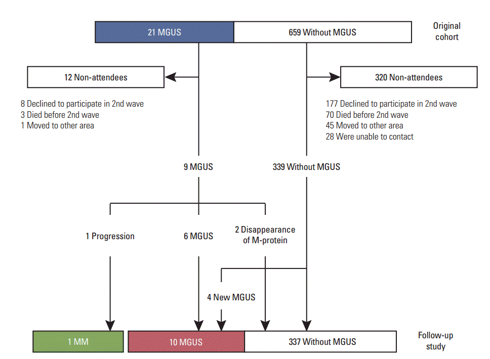
Of the 680 participants (21 with MGUS, 659 without MGUS), 348 (51%) participated in the follow-up study. The median age of the participants was 76 years (range, 70 to 97 years). Ten participants in the follow-up study had MGUS (2.9%; 95% CI, 1.4% to 5.2%) (Table 1, Fig. 1), and the frequency increased with age (odds ratio, 1.6; p for trend, 0.180).
Among the 21 MGUS patients in the initial cohort, nine were followed up (Fig. 1) and six were found to have persistent MGUS. One of these patients had anemia with a persistent M-protein level of 1.4 g/dL, suggestive of progression to multiple myeloma. However, this could not be confirmed by marrow biopsy because of early death immediately after screening. The M-protein disappeared in the remaining two individuals. A new M-protein was detected in four of the 339 participants without MGUS that were followed up.
2. Participant characteristics regarding risk alteration
Table 2 summarizes the characteristics of participants with altered M-protein levels in the follow-up study. Two patients with MGUS in the cohort exhibited the disappearance of M-protein and a normal FLC ratio. Among the four patients with newly developed MGUS, the isotype of monoclonal immunoglobulin was IgG and IgA in three and one patients, respectively, while the immunoglobulin light-chain isotypes were in κ and λ in three and one patients, respectively. An abnormal κ / λ FLC ratio was observed in one of the four new MGUS cases.
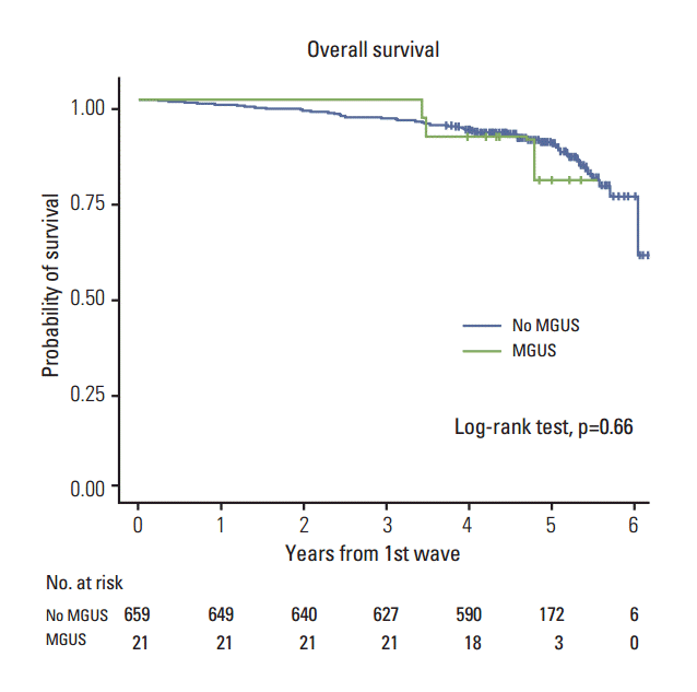
3. Overall survival
A total of 73 deaths (11%) were documented in the 5-year follow-up. Among the 659 participants without MGUS, the most common causes of deaths were malignant solid tumor (n=21, 30%), coronary artery disease (n=8, 11%), cerebral hemorrhage (n=6, 9%), diabetes mellitus (n=4, 6%), and respiratory disease (n=2, 3%). Among the 21 participants with MGUS, three died from diabetes, Parkinsonism, or respiratory disease.
Kaplan-Meier survival analysis revealed no significant difference in survival between participants with and without MGUS detected initially (p=0.66) (Fig. 2).
Discussion
We previously reported the prevalence of MGUS in elderly Koreans (≥ 65 years old) to be 3.3%, which was the first population-based cohort study of MGUS in Korea [6]. The present 5-year follow-up study shows that the overall rate of malignant progression is 5% at 5 years (1 progression/21 MGUS cases), which is consistent with Western studies [4].
The prevalence of MGUS varies by ethnicity, suggesting genetic susceptibility [7,8]. Two large studies from the United States reported that the prevalence of MGUS is 2- to 3-fold greater among African Americans than Caucasians [8,9]. However, there is a paucity of information regarding the incidence of MGUS in Asians [2]. In a large population-based study of atomic bomb survivors in Japan, the overall prevalence of MGUS was 2.4% in those ≥ 50 years of age [10]. However, considering the radiation exposure of residents in Nagasaki, the applicability of these data to the general Asian population is limited.
Kyle et al. [11] reported the prevalence of MGUS to be 3.2% in individuals ≥ 50 years in a population-based study. The present cohort comprised a random sample of elderly people living in a suburb, accurately representing the elderly population (≥ 65 years). Even though the mean age of the present cohort was older than that of the Mayo Clinic cohort, the prevalence of MGUS was considerable. Because there were few MGUS patients in both the initial and follow-up studies, it is not possible to draw a conclusion regarding the rate of progression. However, as the study participants accurately represent the elderly population, we conclude that the observed progression rate at 5 years is 5%, which is similar to that in Western countries. Regardless, this conclusion should be interpreted cautiously.
The M-protein disappeared in two MGUS patients upon follow-up, while the disappearance of M-protein was observed in 0.4% to 4% of MGUS patients in previous studies [4,12]. However, the underlying mechanism of the disappearance of M-protein remains poorly understood. Considering that the stromal component of the bone marrow microenvironment is essential for the development of MGUS [13], the disappearance of M-protein may be related to the dysregulation of various cytokines from non-tumorous cells. It should be noted that we changed the initial screening method from immunofixation using plasma in the initial study to serum protein electrophoresis in the later study. This could have contributed to the decreased sensitivity in the follow-up study. However, we used serum electrophoresis as the standard screening method for MGUS because the amounts of M-protein, which were so small that they can only be detected by immunofixation, have no clinical significance.
Although the risk of MGUS progression at 25 years is 25% to 30%, the prognostic implications of MGUS on survival are not obvious because of the late onset of MGUS and high rate of death from comorbidities [13]. Moreover, none of the 73 deaths observed in the present study were due to hematologic malignancies. Overall, there was no significant difference in survival with respect to the presence of MGUS. While several studies have reported conflicting results regarding the survival of MGUS patients, its absolute impact is expected to be modest in the general population [14,15].
It should be noted that the present study had several limitations. First, almost half of the participants (n=332, 49%) did not participate in the 5-year follow-up evaluation. Among them, 185 participants refused to complete the follow-up evaluation, 73 died during the study period, 46 moved to another area, and contact was lost with 28. Because the KLoSHA involves a cohort of the elderly population (median age of 79) and participants did not receive any intervention, they were not motivated to participate in the follow-up study. Second, bone marrow aspirates were unavailable; therefore, it remains uncertain whether some of the progressive MGUS cases would have been judged as multiple myeloma at baseline. However, the long period between the baseline evaluation and progression to myeloma makes this unlikely. Finally, the presence of clonal plasma cells in the bone marrow was not confirmed in one progressive case of multiple myeloma. However, both the persistent serum monoclonal protein for 5 years and new onset of end-organ damage (i.e., anemia) suggest that the patient had clinically progressed to multiple myeloma.
Conclusion
In conclusion, this population-based cohort study shows that the natural clinical course of MGUS in Korea is similar to that in Western countries. A diagnosis of MGUS was not associated with an increased risk of death in a Korean elderly population. Nevertheless, the large losses to follow-up are a major limitation of the present study.