| Home | E-Submission | Sitemap | Contact Us |  
top_img
Cancer Research and Treatment > Volume 51(4); 2019 > Article
Go, Ko, Lee, Lee, Jeong, Lee, Hong, and Ha: The Use of CD44 Variant 9 and Ki-67 Combination Can Predicts Prognosis Better Than Their Single Use in Early Gastric Cancer

Abstract

Purpose

We previously demonstrated that CD44v9 and Ki-67 played an important role in predicting poor prognosis of early gastric cancer (EGC). However, little is known about combined use of both biomarkers as prognostic biomarker. The present study was performed to investigate the significance of CD44v9 and Ki-67 expression as a combination biomarker for EGC.

Materials and Methods

With tissue microarray for 158 EGC tissues, we performed immunohistochemical staining for CD44v9 and Ki-67. The whole patients were divided into three groups (group A, CD44v9-negative/Ki-67–low; group B, neither group A or C; and group C, CD44v9-positive/Ki-67–high). Its clinical significance was re-analyzed with adjustment via propensity score matching (PSM). For validation, we performed bootstrap resampling.

Results

The median follow-up duration was 90.4 months (range, 3.7 to 120.4 months). In the comparison according to CD44v9/Ki-67 expression, the combined use of the two biomarker clearly separated the three groups by 5-year survival rates (5-YSR, 96.3%, 89.8%, and 76.8% in group A, B, and C, respectively; p=0.009). After PSM, 5-YSR were 97.7% and 76.8% in group A+B and group C, respectively (p=0.002). Multivariable analysis demonstrated that group C had independently poor prognosis (hazard ratio, 9.137; 95% confidence interval, 1.187 to 70.366; p=0.034) compared with group A. Bootstrap resampling internally validated this result (p=0.016).

Conclusion

This study suggests that both positive CD44v9 and high Ki-67 expression are associated with poor prognosis in EGC, and the combined use of these markers provides better prognostic stratification than the single use of them.

Introduction

Notably, gastric cancer (GC) is the third leading cause of cancer-related death worldwide [1] and the second cause of cancer-related death in Korea [2,3]. Histologically, GC has various histologic subtypes and heterogeneity. The cancer heterogeneity can be an obstacle in finding out biomarkers because of its large subgroup where the biomarker does not play a role in predicting prognosis or treatment response, which diminished the effect size [4,5]. Till now, pathologic TNM stage has been still serving as one of the best prognostic markers for GC. However, it is well-known that the prognosis varies in early GC (EGC); some of EGCs shows distant metastasis right after radical resection. Therefore, finding out a new biomarker is essential to predict prognosis of the GC even in EGC.
Recently, we have reported that CD44 variant 9 (CD44v9) and Ki-67 expression can serve as prognostic biomarkers in EGC [4,5]. CD44v9, a variant major adhesion molecule for the extracellular matrix (a surface receptor for hyaluronic acid) [6] is highly expressed at stem cells in mouse gastric tumors [7], and abundantly expressed in epithelial-type carcinomas [8]. It has been implicated in cancer cell adhesion [9], migration [10], and metastasis [11]. In addition, CD44v9 is associated with resistance to chemotherapy or radiotherapy [8] by increasing reduced glutathione through activating cystine-glutamate exchange transporter [12]. Ki-67 protein is widely used as a biomarker for proliferation to determine benign, and malignant tumor, or histologic grade for malignancy [13]. The expression increases in dividing cells and reaches its peak in M phase [14], so the percentage of Ki-67–expressing cells (Ki-67 labeling index) has been used as a standard procedure to assess the proliferative activity of neoplastic cells [15,16]. In our previous study, each of the proteins (CD44v9 and Ki-67) played an important role in predicting poor prognosis of EGC [4,5]. However, in terms of biological aspects for cancer, the functions of the two proteins are different. CD44v9 is associated with stemness, metastasis, and drug resistance [17-21], while Ki-67 is proliferation and invasion [4,22]. We hypothesized that synergism is expected when the two biomarkers reflecting different activities are used together. In addition, little is known about whether the combined use of the two biomarkers together help to predict better prognosis of EGC than the single biomarker. Therefore, we conducted this study to determine whether the combined use of CD44v9 and Ki-67 expression serves as a better prognostic biomarker in EGC than does the use of single one, either CD44v9 or Ki-67 expression.

Materials and Methods

1. Clinical specimens and patients

Tissue microarray samples were used; they contained 158 EGC and 162 advanced GC (AGC) tissues from the patients with GC who had received radical gastric resection from 1999 to 2007. Tissue microarray samples were made as previously described [5]. Each tissue core contained the invasive front of GC.

2. Immunohistochemical staining

Immunohistochemical (IHC) staining was performed as previously described [4,5]. Briefly, a 4-μm-thick paraffin-embedded tissue microarray on each slide was treated for epitope retrieval. The primary monoclonal antibodies specific to CD44v9 (kindly provided by Dr. Hideyuki Saya, diluted 1:10,000 to 0.1 μg/mL; Keio University School of Medicine, Tokyo, Japan), and Ki-67 (1:2,000, DAKO, Tokyo, Japan) were used. 3,3′-Diaminobenzidine was used for color development and counterstained with hematoxylin. The IHC staining was performed by an automatic staining machine (BenchMark XT, Ventana, Tucson, AZ).
Antigen expression was scored by pathologists with the following guideline and blindness to the clinical data of the patients. The representative figures of CD44v9 and Ki-67 IHC staining are shown in S1 Fig. For CD44v9, the staining intensity was considered as negative when less than 5% of tumor cells were stained, and positive when 5% of tumor cells or more were stained. For Ki-67, various cut-off values have been proposed in previous studies for GC ranging from 1% to 53% [22]. Because there is no established cut-off value for Ki-67, we used our previous definition that the staining intensity was interpreted as low when less than 20% of tumor cells were stained, and high when 20% of tumor cells or more were stained [4]. The whole patients were divided into three groups according to CD44v9 and Ki-67 expression for analysis (group A, CD44v9-negative/Ki-67–low; group B, neither group A or C; and group C, CD44v9-positive/Ki-67–high). Histological type and tumor stage were classified according to the World Health Organization (WHO) [23] and Lauren classification [24], and the 8th edition of American Joint Committee on Cancer TNM system [25], respectively.

3. Statistical analysis

All statistical analyses were performed by STATA ver. 14.0 software (College Station, TX) and R ver. 3.4 software (R Foundation for Statistical Computing, Vienna, Austria). A two-sided p-value of < 0.05 was considered statistically significant. For categorical variables, chi-square, chi-square for trend, and Fisher exact tests were used and Mann-Whitney U and Kruskal-Wallis tests for continuous variables. The overall survival (OS) was defined as the time from surgery to death of any cause or last follow-up. Kaplan-Meier method was used for survival plot and the difference was analyzed by log-rank test. To identify independent prognostic factors, we used Cox regression model, where all the variables moderately associated with survival (p < 0.10) in the univariate analyses were included. The final Cox model was internally validated by bootstrap resampling (1,000 replications). To compare the discriminatory power between CD44v9/Ki-67 and either CD44v9 or Ki-67 alone, the c-indices of each Cox model including these markers were calculated. A c-index of 0.5 indicates random prediction, while a c-index of 1.0 means perfect predictability. Additionally, we performed propensity score matching (PSM) between group A+B and group C to reduce selection bias. Age, sex, and potential predictors for CD44v9/Ki-67 expression having p < 0.05 were included in PSM.

4. Ethical statement

Authors were permitted to use these samples for this study by Institutional Review Board of Gyeongsang National University Hospital (GNUHIRB-2009-19), which waived the requirement for informed consent because of the retrospective nature of the study.

Results

1. Baseline characteristics of EGC patients

Baseline characteristics of 158 EGC patients are presented in Table 1, showing that the median age at the time of surgery was 65 years (range, 26 to 85 years) with a male predominance (n=101, 63.9%). The most frequently performed surgery was subtotal gastrectomy (n=129, 81.7%). Fifteen lymph node or more were excised in 122 patients (77.2%). All but five patients (96.8%) had stage I disease. The histologic grade according to the WHO classification was evenly distributed, whereas the intestinal-type (n=123, 77.9%) was observed to be predominant according to the Lauren classification. No significant differences between groups have been observed in demographics, surgery type, location, lymph node metastasis, stage, and tumor size. However, the histologic subtypes known to be associated with better prognosis in GC (well-differentiated [W/D] histology and intestinal type) were more frequently distributed in the following order of groups C, B, and A (WHO classification, p=0.004; Lauren classification, p=0.007).

2. The role of CD44v9/Ki-67 as a prognostic biomarker in EGC

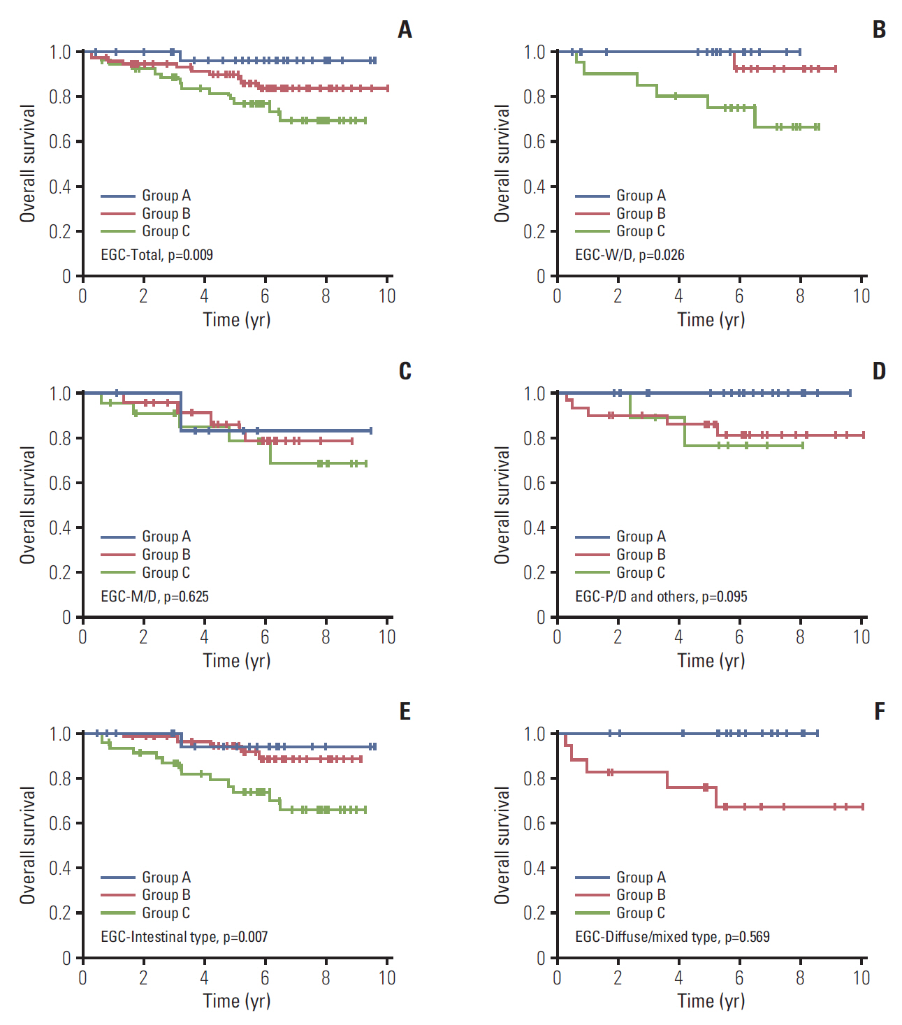
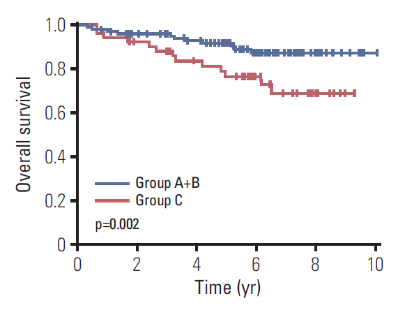
The median follow-up duration was 90.4 months (range, 3.7 to 120.4 months). In all EGC patients, 5-year survival rate (5-YSR) was 86.7% (95% confidence interval [CI], 79.8 to 91.3). Based on CD44v9/Ki-67 expression, 5-YSR were 96.3%, 89.8%, and 76.8% in group A, B, and C, respectively (p=0.009) (Fig. 1A). In subgroup analyses, it was found that CD44v9/Ki-67 expression played a role in predicting prognosis in W/D histology of EGC (p=0.026), but not in other moderately-differentiated (M/D) and poorly-differentiated (P/D) histology of EGC (p=0.625 and p=0.095, respectively) (Fig. 1B-D). When CD44-v9/Ki-67 served as a prognostic factor in intestinal type of EGC (p=0.007) (Fig. 1E), but not in diffuse/mixed type (p= 0.569) (Fig. 1F). After PSM between group A+B and group C (n=106), there were no longer significant differences in the patients’ characteristics (Table 2, S2 Fig.). The 5-YSR adjusted with PSM were 97.7% and 76.8% in group A+B and group C, respectively (p=0.002) (Fig. 2).
On univariate analyses, age ≥ 65, male sex, upper third location, lymph node metastasis, and group C were detected as potential poor prognostic factors for OS. Among them, male sex (hazard ratio [HR], 3.465; 95% CI, 1.135 to 10.584; p=0.029) and group C (HR, 9.137; 95% CI, 1.187 to 70.366; p=0.034) were independent prognostic factors for OS. Bootstrap resampling internally validated this result (p=0.016) (Table 3). The c-indices of multivariate Cox regression models including CD44v9/Ki-67, CD44v9, and Ki-67 variables were 0.75 (95% CI, 0.67 to 0.83), 0.71 (95% CI, 0.61 to 0.81), and 0.72 (95% CI, 0.63 to 0.81), respectively. These findings suggest that combined use of both the biomarkers (CD44v9/Ki-67) serves better as a prognostic biomarker for EGC than the use of single biomarker, that is–either CD44v9 or Ki-67.

3. The role of CD44v9/Ki-67 as a prognostic biomarker in AGC

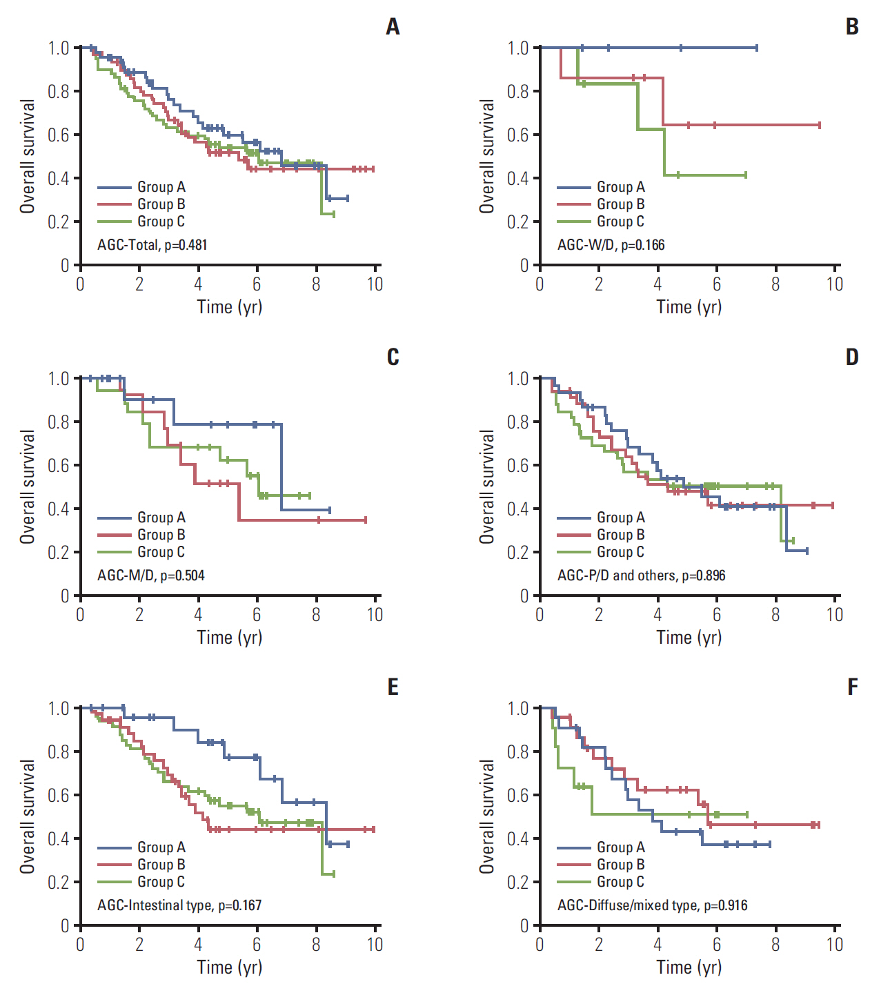
Next, we assessed the role of CD44v9/Ki-67 in AGC patients (n=162). There were no differences in baseline characteristics except age and Lauren classification (S3 Table). It was revealed that CD44v9/Ki-67 did not serve as a prognostic factor in this population (p=0.481) (Fig. 3A). In subgroup analyses, it was found that CD44v9/Ki-67 expression did not serve as a prognostic factor in W/D histology (p=0.166) as well as in M/D and P/D histology (p=0.504 and p=0.896, respectively) (Fig. 3B-D). Similarly, there was no prognostic value of CD44v9/Ki-67 expression in AGC regardless of Lauren classification (Fig. 3E and F). These findings support that CD44v9/Ki-67 played an important role in predicting prognosis in EGC, but not in AGC.

Discussion

This study was designed to determine whether the two biomarkers, CD44v9 and Ki-67 serve better as a prognostic factor in EGC than either any one of them. Hence, we compared survivals among the three groups (negative/low, negative/high or positive/low, and positive/high for CD44v9 and Ki-67 expression) in EGC. It was found that the groups of both up-regulated expression of CD44v9 and Ki-67 showed the worst survival results among the three groups, and validated the results with bootstrap resampling.
The prognosis of cancer is largely determined by cancer metastasis. Metastasis is determined by the activity of various factors such as tumor growth, tissue adhesion, neovascularization, immune evasion, and drug resistance. Aggressiveness of tumor is always challenging and could be represented by the rapid tumor cell proliferation and metastasis capability in cancer. In search of biomarkers for the aggressiveness or metastasis, it has been reported that CD44v9 serves as stem cell, and metastasis biomarker [17-21], and Ki-67 represents as proliferation and invasion biomarker [4,14,22]. Therefore, the use of a biomarker to evaluate each of these factors can be more accurate in assessing the prognosis than using a biomarker with a monopoly. The synergism is expected when the two biomarkers reflecting different activities are used together for prognosis. This assumption was confirmed in this study by showing that the EGC expressing both high Ki-67 (proliferation biomarker) and positive CD44v9 (stem cell, and metastasis biomarker) are with the worst outcome and that the Cox regression model including CD44v9/Ki-67 expression showed a higher discriminating ability for survival compared with those including either CD44v9 or Ki-67 expression alone. In addition, multigene assays have been widely used as a guideline for adjuvant chemotherapy for early breast cancer treatment [26,27]. This study also confirm that the combined use of two biomarkers serves as a significant prognostic factors only in EGC but not in AGC although the combined use better predicts prognosis of GC than single use.
The limitation in this investigation is that we could not clearly demonstrate the difference in role of CD44v9 and Ki-67 combination as a prognostic biomarker in M/D, and P/D histologic subtype of EGC, which is presume to be due to the small sample size. Previous studies reported that Ki-67 tended to better reflect the prognosis in the W/D histologic type of EGC [4]. Moreover, histological observation studies have revealed that, W/D type is predominant in EGC of the elderly, while in AGC, predominant is mixed type which is composed of W/D type in the superficial area, and P/D type in the deeper area [28,29]. These findings suggest that majority of intestinal type of AGCs are principally W/D adenocarcinomas which have progressed to P/D adenocarcinomas as EGC progress to AGC. Taking into account this fact, M/D and P/D histological subtype could be more heterogeneous than W/D type. In this context, there is a possibility that CD44v9 and Ki-67 combination serves less as a prognostic biomarker in M/D, and P/D histologic subtype of EGC than in W/D histologic subtype of EGC. Therefore, further larger research is warranted at this point.
The merit of this study is that we have applied two biomarkers with different function at the same staged cancer, which is making it more chance to avoid heterogeneity. Previous many studies investigated role of each of the biomarkers Ki-67, standard or variant of CD44 in the EGC or for whole GC group including AGC [4,5,30-32]. In these studies, the results in EGC was clear than the others. Secondly, we had performed propensity-score matching analysis, and validated the final results with bootstrap resampling to eliminate hidden biases. The last point to emphasize is that this study indicated that the prognosis of the patients with cancer expressing both positive CD44v9 and high Ki-67 was as poor as those with AGC, which suggests that these patients need further treatment to reduce relapse of GC.
In conclusion, these results suggest that both positive CD44v9 and high Ki-67 expression are associated with poor prognosis in EGC, and the combined use of these markers provides better prognostic stratification than the single use of them.

Electronic Supplementary Material

Supplementary materials are available at Cancer Research and Treatment website (https://www.e-crt.org).

Conflicts of Interest

Conflict of interest relevant to this article was not reported.

Acknowledgments

This study was supported by a grant of the National R&D Program for Cancer Control, Ministry for Health, Welfare and Family Affairs, Republic of Korea (0820050), in part by a grant from the Korean Cancer Research Institute. We really appreciate Dr. Suchismita Raha’s advice and English editing in writing the manuscripts.

Fig. 1.
Kaplan-Meier curves for overall survival according to CD44v9 and Ki-67 expression in early gastric cancer (EGC) patients. (A) Total. (B) Well-differentiated (W/D). (C) Moderately-differentiated (M/D). (D) Poorly-differentiated (P/D) and others. (E) Intestinal type. (F) Diffuse/mixed type. Others indicate undifferentiated and mucinous adenocarcinoma, and signet ring cell carcinoma.
crt-2018-663f1.jpg
Fig. 2.
Kaplan-Meier curves for overall survival according to CD44v9 and Ki-67 expression in early gastric cancer after propensity score matching.
crt-2018-663f2.jpg
Fig. 3.
Kaplan-Meier curves for overall survival according to CD44v9 and Ki-67 expression in advanced gastric cancer (AGC) patients. (A) Total. (B) Well-differentiated (W/D). (C) Moderately-differentiated (M/D). (D) Poorly-differentiated (P/D) and others. (E) Intestinal type. (F) Diffuse/mixed type. Others indicate undifferentiated and mucinous adenocarcinoma, and signet ring cell carcinoma.
crt-2018-663f3.jpg
Table 1.
Baseline characteristics of early gastric cancer patients
Characteristic Group A (n=32) Group B (n=73) Group C (n=53) p-value
Age (yr) 61 (35-78) 65 (32-78) 66 (26-85) 0.183
Sex
 Male 24 (75.0) 48 (65.8) 29 (54.7) 0.054
 Female 8 (25.0) 25 (34.3) 24 (45.3)
Location
 Upper 2 (6.3) 5 (6.9) 3 (5.7) 0.470
 Middle 8 (25.0) 18 (24.7) 10 (18.9)
 Lower 22 (68.8) 50 (68.5) 40 (75.5)
Operation
 Subtotal gastrectomy 25 (78.1) 58 (79.5) 46 (86.8) 0.276
 Total gastrectomy 4 (12.5) 11 (15.1) 4 (7.6)
 Proximal gastrectomy 2 (6.3) 4 (5.5) 2 (3.8)
 Wedge resection 1 (3.1) 0 1 (1.9)
No. of lymph nodes dissection
 < 15 8 (25.0) 13 (17.8) 15 (28.3) 0.561
 ≥ 15 24 (75.0) 60 (82.2) 38 (71.7)
Regional lymph node metastasis
 Negative 30 (93.8) 65 (89.0) 47 (88.7) 0.495
 Positive 2 (6.3) 8 (11.0) 6 (11.3)
TNM stage
 I 31 (96.9) 72 (98.6) 50 (94.3) 0.402
 II 1 (3.1) 1 (1.4) 3 (5.7)
Tumor size (cm) 2.2 (1.0-5.5) 2.5 (1.0-11.0) 2.5 (0.5-7.0) 0.354
WHO classification
 Well-differentiated 8 (25.0) 20 (27.4) 21 (39.6) 0.004
 Moderately-differentiated 7 (21.9) 24 (32.9) 22 (41.5)
 Poorly-differentiated and othersa) 17 (53.1) 29 (39.7) 10 (18.9)
Lauren classification
 Intestinal 20 (62.5) 56 (76.7) 47 (88.7) 0.007
 Diffuse 10 (31.3) 15 (20.6) 3 (5.7)
 Mixed 2 (6.3) 2 (2.7) 3 (5.7)

Values are presented as median (range) or number (%). WHO, World Health Organization.

a) Undifferentiated and mucinous adenocarcinoma, and signet-ring cell carcinoma were included.

Table 2.
Baseline characteristics of early gastric cancer patients after propensity score matching (PSM)
Group A+B (before PSM) (n=105) Group A+B (after PSM) (n=53) Group C (n=53) p-value (group A+B vs. C before PSM) p-value (group A+B vs. C after PSM)
Age (yr) 64 (32-78) 67 (32-77) 66 (26-85) 0.133 0.613
Sex
 Male 72 (68.6) 32 (60.4) 29 (54.7) 0.087 0.556
 Female 33 (31.4) 21 (39.6) 24 (45.3)
Location
 Upper 7 (6.7) 2 (3.8) 3 (5.7) 0.660 0.821
 Middle 26 (24.8) 12 (22.6) 10 (18.9)
 Lower 72 (68.6) 39 (73.6) 40 (75.5)
Operation
 Subtotal gastrectomy 83 (79.1) 42 (79.3) 46 (86.8) 0.553 0.753
 Total gastrectomy 15 (14.3) 7 (13.2) 4 (7.6)
 Proximal gastrectomy 6 (5.7) 3 (5.7) 2 (3.8)
 Wedge resection 1 (1.0) 1 (1.9) 1 (1.9)
No. of lymph nodes dissection
 < 15 21 (20.0) 12 (22.6) 15 (28.3) 0.240 0.504
 ≥ 15 84 (80.0) 41 (77.4) 38 (71.7)
Regional lymph node metastasis
 Negative 95 (90.5) 47 (88.7) 47 (88.7) 0.724 > 0.990
 Positive 10 (9.5) 6 (11.3) 6 (11.3)
TNM stage
 I 103 (98.1) 52 (98.1) 50 (94.3) 0.335 0.618
 II 2 (1.9) 1 (1.9) 3 (5.7)
Tumor size (cm) 2.5 (1.0-11.0) 2.5 (1.0-11.0) 2.5 (0.5-7.0) 0.368 0.484
WHO classification
 Well-differentiated 28 (26.7) 25 (47.2) 21 (39.6) 0.008 0.478
 Moderately-differentiated 31 (29.5) 16 (30.2) 22 (41.5)
 Poorly-differentiated and othersa) 46 (43.8) 12 (22.6) 10 (18.9)
Lauren classification
 Intestinal 76 (72.4) 47 (88.7) 47 (88.7) 0.018 0.472
 Diffuse 25 (23.8) 5 (9.4) 3 (5.7)
 Mixed 4 (3.8) 1 (1.9) 3 (5.7)

Values are presented as median (range) or number (%). WHO, World Health Organization.

a) Undifferentiated and mucinous adenocarcinoma, and signet-ring cell carcinoma were included.

Table 3.
Cox regression for overall survival in early gastric cancer patients
Univariate
Multivariate
HR 95% CI p-value HR 95% CI p-value p-value (bootstrap)
Age (≥ 65 yr vs. < 65 yr) 2.178 0.932-5.092 0.072 1.913 0.794-4.607 0.148 0.153
Sex (male vs. female) 2.751 0.940-8.049 0.065 3.465 1.135-10.584 0.029 0.019
Location (upper vs. middle/lower) 2.999 1.023-8.793 0.045 1.560 0.461-5.274 0.475 0.540
Operation (subtotal gastrectomy vs. others) 1.164 0.435-3.119 0.762 - - - -
No. of lymph nodes dissection (< 15 vs. ≥ 15) 1.814 0.776-4.243 0.169 - - - -
Regional lymph node metastasis (negative vs. positive) 2.962 1.004-8.744 0.049 2.609 0.744-9.143 0.134 0.183
Tumor size (≥ 4 cm vs. < 4 cm) 0.605 0.226-1.620 0.317 - - - -
WHO classification
 Well-differentiated Reference
 Moderately-differentiated 1.547 0.587-4.075 0.377 - - - -
 Poorly-differentiated and othersa) 0.923 0.324-2.633 0.881 - - - -
Lauren classification (diffuse/mixed vs. intestinal) 0.920 0.344-2.465 0.869 - - - -
CD44v9 and Ki-67
 Group A Reference Reference
 Group B 4.510 0.577-35.232 0.151 4.070 0.515-32.194 0.183 0.092
 Group C 8.475 1.109-64.795 0.039 9.137 1.187-70.366 0.034 0.016

HR, hazard ratio; CI, confidence interval; WHO, World Health Organization.

a) Undifferentiated and mucinous adenocarcinoma, and signet-ring cell carcinoma were included.

References

1. Siegel RL, Miller KD, Jemal A. Cancer statistics, 2016. CA Cancer J Clin. 2016;66:7–30.
crossref pmid
2. Song HN, Go SI, Lee WS, Kim Y, Choi HJ, Lee US, et al. Population-based regional cancer incidence in Korea: comparison between urban and rural areas. Cancer Res Treat. 2016;48:789–97.
crossref pmid pdf
3. Oh CM, Won YJ, Jung KW, Kong HJ, Cho H, Lee JK, et al. Cancer statistics in Korea: incidence, mortality, survival, and prevalence in 2013. Cancer Res Treat. 2016;48:436–50.
crossref pmid pmc pdf
4. Ko GH, Go SI, Lee WS, Lee JH, Jeong SH, Lee YJ, et al. Prognostic impact of Ki-67 in patients with gastric cancer-the importance of depth of invasion and histologic differentiation. Medicine (Baltimore). 2017;96:e7181
crossref pmid pmc
5. Go SI, Ko GH, Lee WS, Kim RB, Lee JH, Jeong SH, et al. CD44 variant 9 serves as a poor prognostic marker in early gastric cancer, but not in advanced gastric cancer. Cancer Res Treat. 2016;48:142–52.
crossref pmid pdf
6. Aruffo A, Stamenkovic I, Melnick M, Underhill CB, Seed B. CD44 is the principal cell surface receptor for hyaluronate. Cell. 1990;61:1303–13.
crossref pmid
7. Ishimoto T, Oshima H, Oshima M, Kai K, Torii R, Masuko T, et al. CD44+ slow-cycling tumor cell expansion is triggered by cooperative actions of Wnt and prostaglandin E2 in gastric tumorigenesis. Cancer Sci. 2010;101:673–8.
crossref pmid
8. Diehn M, Cho RW, Lobo NA, Kalisky T, Dorie MJ, Kulp AN, et al. Association of reactive oxygen species levels and radioresistance in cancer stem cells. Nature. 2009;458:780–3.
crossref pmid pmc pdf
9. Culty M, Miyake K, Kincade PW, Sikorski E, Butcher EC, Underhill C. The hyaluronate receptor is a member of the CD44 (H-CAM) family of cell surface glycoproteins. J Cell Biol. 1990;111(6 Pt 1):2765–74.
crossref pmid pmc
10. Thomas L, Byers HR, Vink J, Stamenkovic I. CD44H regulates tumor cell migration on hyaluronate-coated substrate. J Cell Biol. 1992;118:971–7.
crossref pmid pmc
11. Gunthert U, Hofmann M, Rudy W, Reber S, Zoller M, Haussmann I, et al. A new variant of glycoprotein CD44 confers metastatic potential to rat carcinoma cells. Cell. 1991;65:13–24.
crossref pmid
12. Ishimoto T, Nagano O, Yae T, Tamada M, Motohara T, Oshima H, et al. CD44 variant regulates redox status in cancer cells by stabilizing the xCT subunit of system xc(-) and thereby promotes tumor growth. Cancer Cell. 2011;19:387–400.
crossref pmid
13. Scholzen T, Gerdes J. The Ki-67 protein: from the known and the unknown. J Cell Physiol. 2000;182:311–22.
crossref pmid
14. Gerdes J, Lemke H, Baisch H, Wacker HH, Schwab U, Stein H. Cell cycle analysis of a cell proliferation-associated human nuclear antigen defined by the monoclonal antibody Ki-67. J Immunol. 1984;133:1710–5.
crossref pmid pdf
15. Deshmukh P, Ramsey L, Garewal HS. Ki-67 labeling index is a more reliable measure of solid tumor proliferative activity than tritiated thymidine labeling. Am J Clin Pathol. 1990;94:192–5.
crossref pmid pdf
16. Cher ML, Chew K, Rosenau W, Carroll PR. Cellular proliferation in prostatic adenocarcinoma as assessed by bromodeoxyuridine uptake and Ki-67 and PCNA expression. Prostate. 1995;26:87–93.
crossref pmid
17. Al-Hajj M, Wicha MS, Benito-Hernandez A, Morrison SJ, Clarke MF. Prospective identification of tumorigenic breast cancer cells. Proc Natl Acad Sci U S A. 2003;100:3983–8.
crossref pmid pmc
18. Dalerba P, Dylla SJ, Park IK, Liu R, Wang X, Cho RW, et al. Phenotypic characterization of human colorectal cancer stem cells. Proc Natl Acad Sci U S A. 2007;104:10158–63.
crossref pmid pmc
19. Zavros Y. Initiation and maintenance of gastric cancer: a focus on CD44 variant isoforms and cancer stem cells. Cell Mol Gastroenterol Hepatol. 2017;4:55–63.
crossref pmid pmc
20. Singh SR. Gastric cancer stem cells: a novel therapeutic target. Cancer Lett. 2013;338:110–9.
crossref pmid pmc
21. Kodama H, Murata S, Ishida M, Yamamoto H, Yamaguchi T, Kaida S, et al. Prognostic impact of CD44-positive cancer stem-like cells at the invasive front of gastric cancer. Br J Cancer. 2017;116:186–94.
crossref pmid pdf
22. Luo G, Hu Y, Zhang Z, Wang P, Luo Z, Lin J, et al. Clinicopathologic significance and prognostic value of Ki-67 expression in patients with gastric cancer: a meta-analysis. Oncotarget. 2017;8:50273–83.
crossref pmid pmc
23. Hu B, El Hajj N, Sittler S, Lammert N, Barnes R, Meloni-Ehrig A. Gastric cancer: classification, histology and application of molecular pathology. J Gastrointest Oncol. 2012;3:251–61.
pmid pmc
24. Lauren P. The two histological main types of gastric carcinoma: diffuse and so-called intestinal-type carcinoma: an attempt at a histo-clinical classification. Acta Pathol Microbiol Scand. 1965;64:31–49.
crossref pmid
25. Amin MB, Edge S, Greene F, Byrd DR, Brookland RK, Washington MK, et al. AJCC cancer staging manual. 8th edNew York, NY: Springer; 2016.

26. Jerzak KJ, Pritchard KI. The 21-gene recurrence score assay in node-negative early breast cancer: prognostic, predictive or presumptuous? Eur J Cancer. 2016;68:173–5.
crossref pmid
27. Sestak I, Buus R, Cuzick J, Dubsky P, Kronenwett R, Denkert C, et al. Comparison of the performance of 6 prognostic signatures for estrogen receptor-positive breast cancer: a secondary analysis of a randomized clinical trial. JAMA Oncol. 2018;4:545–53.
crossref pmid pmc pdf
28. Inoshita N, Yanagisawa A, Arai T, Kitagawa T, Hirokawa K, Kato Y. Pathological characteristics of gastric carcinomas in the very old. Jpn J Cancer Res. 1998;89:1087–92.
crossref pmid pmc
29. Arai T, Esaki Y, Inoshita N, Sawabe M, Kasahara I, Kuroiwa K, et al. Pathologic characteristics of gastric cancer in the elderly: a retrospective study of 994 surgical patients. Gastric Cancer. 2004;7:154–9.
crossref pmid pmc pdf
30. Hassani Joutei HA, Marchoudi N, Sadaoui I, Mahfoud W, Haddad F, Fechtali T, et al. Ki-67 proliferative index in gastric cancer: a useful prognostic marker? Anticancer Res. 2014;34:5806.

31. Costa L, Fradique AC, Pupo A, Quaresma L, Cabrita F, Da Silva G, et al. Ki 67 showed statistical prognostic significance in intestinal type of gastric cancer. Ann Oncol. 2013;24(Suppl 4):iv59.
crossref pdf
32. Fang M, Wu J, Lai X, Ai H, Tao Y, Zhu B, et al. CD44 and CD44v6 are correlated with gastric cancer progression and poor patient prognosis: evidence from 42 studies. Cell Physiol Biochem. 2016;40:567–78.
crossref pmid
TOOLS
PDF Links  PDF Links
PubReader  PubReader
ePub Link  ePub Link
XML Download  XML Download
Full text via DOI  Full text via DOI
Download Citation  Download Citation
Supplement  Supplement1
Supplement  Supplement2
Supplement  Supplement3
  Print
Share:      
METRICS
12
Web of Science
10
Crossref
13
Scopus
7,135
View
173
Download
Related article
Editorial Office
Korean Cancer Association
Room 1824, Gwanghwamun Officia
92 Saemunan-ro, Jongno-gu, Seoul 03186, Korea
TEL: +82-2-3276-2410   FAX: +82-2-792-1410   E-mail: journal@cancer.or.kr
About |  Browse Articles |  Current Issue |  For Authors and Reviewers
Copyright © Korean Cancer Association.                 Developed in M2PI