Introduction
Choroidal melanomas are the most common primary intraocular malignancies in adults. However, choroidal melanoma predominantly affects Caucasians, while it is very rare in Asian populations. Despite the low incidence rates in Asian populations, the tumors usually affect younger patients, tend to be larger, and present a high number of epitheloid cell types, resulting in a poorer prognosis in Asian patients than in Caucasians [1].
Until the introduction of plaque brachytherapy using iodine 125 (125I) in the 1960s, enucleation was the standard treatment for patients with choroidal melanomas. Although the most important treatment goal is survival free of disease recurrence, both patients and clinicians focus on preservation of function and cosmetic appearance as well. The Collaborative Ocular Melanoma Study confirmed the efficacy of plaque brachytherapy in a multicenter randomized trial including 1,317 patients, which did not reveal any inferiority of 125I brachytherapy to enucleation, showing comparable 5-year overall survival (OS) (81% vs. 82%) and 5-year cancer-specific survival (89% vs. 91%) rates [2,3]. Moreover, 125I brachytherapy did pose any additional risk of loss of visual acuity in the fellow eye for at least 10 years after treatment for choroidal melanoma [4]. Today, plaque brachytherapy is a major modality for the management of choroidal melanomas because it is considered a reasonable eye-preserving alternative to enucleation. Although the benefits of plaque brachytherapy may be reduced by impaired vision resulting from radiation toxicity, this treatment modality has major functional and cosmetic advantages.
Since its introduction, ruthenium-106 (106Ru) brachytherapy has been increasingly used for the treatment of small- to medium-sized choroidal melanomas [5-7]. A 106Ru plaque providing 1,000 Gy to the scleral surface conveys less than 100 Gy to a distance over 7 mm from the base, giving an insufficient radiation dose to the apex of large tumors [8,9]. Insufficient radiation to the apex could reduce the local tumor control rate and increase the risk of recurrence, as well as metastases and mortality. Several studies have reported outcomes of the combination of the brachytherapy and transpupillary thermotherapy (TTT) [10,11]. This combination therapy was introduced to enable eye-preservation for patients with tumors larger than 5 mm, although it has also been used to lessen the radiation dose for smaller tumors to reduce the risk of radiation-related toxicities. Moreover, patients with insufficient tumor regression after brachytherapy or with recurrent tumors can be re-treated with TTT.
Before the introduction of 106Ru plaques (Eckert & Ziegler BEBIG, Berlin, Germany) to our institution in October 2006, Gamma knife surgery (GKS) using a Leksell γ-knife (Elekta Instruments AB, Stockholm, Sweden) was the only eye-sparing treatment for choroidal melanomas available in Korea. Since 2006, we have primarily performed brachytherapy with additional local treatment, including TTT and local excision, as eye-preserving therapy for choroidal melanomas. This study was conducted to report the early clinical outcomes of this eye-preserving treatment strategy for variable-sized choroidal melanomas in Korean patients.
Materials and Methods
1. Patient selection
We identified 93 consecutive patients who were diagnosed with uveal malignant melanomas and treated with 106Ru brachytherapy between 2006 and 2012 in our institution. Patients with iris and ciliary body melanomas were excluded (n=5) from this study. Because 106Ru plaque therapy is generally considered to treat tumors less than 6-7 mm in height, our patients were divided into a large group (tumor height ≥ 6 mm) and a small group (tumor height < 6 mm) according to their tumor height, as measured by ultrasonography.
2. Treatment methods
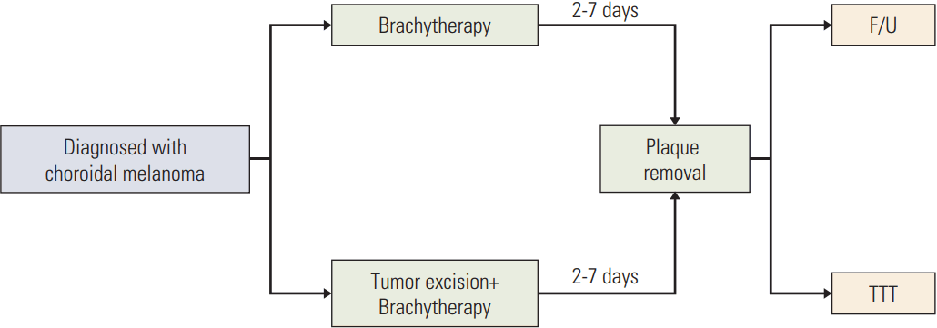
The treatment flow diagram is presented in Fig. 1. The treatment was intended to deliver 85-95 Gy to the apex of the tumor. However, owing to rapid radiation dose fall-off of 106Ru, 80% of the dose had already been absorbed at 5 mm from the applicator. Thus, the dose to the apex is generally considered insufficient for treatment of large tumors. The patients were informed of this limitation, but some patients preferred brachytherapy anyway in an effort to preserve their eyes.
The three most frequently used applicators have diameters of 15.3, 17.9, and 20.2 mm with active diameters of 13.5, 15.8, and 18 mm, respectively. Applicators for the treatment of juxtapapillary tumors (those located immediately adjacent to the optic nerve) have a section cutout for the optic nerve.
Some patients treated with brachytherapy received additional TTT and/or local excision. Local excision of the tumor was conducted before brachytherapy on the same day as the brachytherapy, which was delivered as an adjuvant treatment. Planned TTT was performed with an adjuvant goal after brachytherapy in patients who did not undergo local excision. Unplanned TTT was performed if tumor regression during follow-up was not sufficient after brachytherapy, regardless of initial tumor size. The first planned TTT procedure was performed at the time of plaque removal using an infrared diode laser and a slit-lamp delivery system. During the follow-up period, additional TTT procedures were performed in patients in an outpatient setting at intervals of 3 months.
Enucleation was generally recommended for patients with large tumors (height ≥ 6 mm) because 106Ru brachytherapy is not suitable for such tumors. However, for patients who refused enucleation, local excision of the tumor was performed to reduce the height of the apex. If the tumor was mainly located near the ciliary body, a transscleral tumor resection was performed, while if the tumor had a posterior location and a basal diameter of less than 15 mm, endoresection with pars plana vitrectomy was performed.
3. Evaluation of outcomes
The primary end-point was the eye-preservation rate (free from enucleation). In addition, local control, OS and the rate of distant metastasis were assessed. Survival was calculated from the date of the insertion of the eye plaque to any event. Patients lost to follow-up were censored. Late toxicity was evaluated according to the Radiation Therapy Oncology Group scale.
Tumor response was evaluated with fundoscopy and ultrasonography, with a flat scar or a regressed lesion not showing any signs of tumor activity representing tumor control. Local recurrence was defined as signs of tumor activity on fluorescein angiography, or documented tumor growth of more than 20% of the initial diameter or thickness (Response Evaluation Criteria in Solid Tumors criteria). Metastases were detected through routine follow-up investigations (liver ultrasonography and blood chemistry tests) or using 18F-fluorodeoxyglucose positron emission tomography.
To assess the toxicity of treatment, regular follow-ups for visual acuity and other ophthalmologic evaluations were performed, in which complications such as retinopathy, maculopathy, optic neuropathy, retinal hemorrhage, and exudative retinal detachment were evaluated. In this study, only issues that needed further treatment were defined as complications. Vision was assessed using the World Health Organization (WHO) criteria, with low vision defined as a visual acuity less than 1/3 and a vision < 0.1 considered legal blindness.
4. Statistical analysis
The survival curves were estimated using the Kaplan-Meier method, and comparisons between the two patients groups were made with the log-rank test. Significant variables in univariate analysis (p < 0.05) were entered into a multivariable Cox regression model to identify independent predictors of eye-preservation rate, distant metastasis, and OS. p < 0.05 were considered significant. Statistical analyses were performed using SPSS ver. 20.0.0 (SPSS Inc., Chicago, IL).
Results
1. Patient and treatment characteristics
Between 2006 and 2012, 93 patients with choroidal melanomas were treated with 106Ru brachytherapy. Five patients with iris and ciliary body tumors were excluded, leaving 88 patients in the analysis. The median follow-up period was 30 months (range, 2 to 79 months). The patients and treatment characteristics are summarized in Table 1. The median age was 49 years (range, 19 to 82 years). The initial visual acuity in the affected eye was ≥ 0.10 in 71% (n=63) and ≥ 0.9 in 16% (n=14) of patients. Twenty-five (29%) patients were diagnosed with legal blindness (< 0.1) prior to treatment and 10 patients (11%) had visual impairment ((< 20/60-20/200, WHO definition). The median basal tumor diameter was 11.4 mm (range, 2.6 to 17.3 mm), and the median tumor height was 6.8 mm (range, 1.7 to 13.7 mm). The number of patients with a tumor height ≥ 6 mm (large group) was 50 (57%), and there were 38 (43%) patients in the small group.
Combined therapy was used in the treatment of 75% (n=65) of the patients. In the large group (tumor height ≥ 6 mm), 96% (n=48) of the patients received combined local therapy, while in the small group, 44.7% (n=17) of the patients were treated with combined treatment. The radiation dose was delivered to 2-6 mm from the outer surface of the sclera for 47.7% (n=42) of the entire sample, based on the extent of local excision and use of TTT. For some of the patients who were treated with TTT alone as an additional local therapy and for all patients who did not receive additional treatment, the tumor apex was the prescription point.
Some patients (n=15, 39.5%) in the small group also received additional TTT when tumor regression was deemed insufficient after brachytherapy, while two patients received local excision before brachytherapy. One patient experienced rupture of the tumor during vitrectomy and eventually received internal choroidectomy. The other patient, who had a tumor with a size of 12.5 mm (basal diameter)×5.9 mm (tumor height), was included in the small group in this study. However, he received local excision based on the decision of his physicians.
2. Local control, distant metastasis, and OS
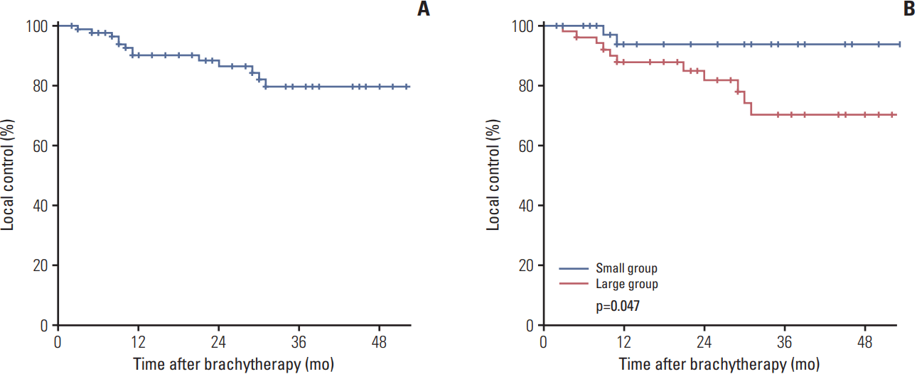
Local progression was diagnosed in 13 patients. The 3-year local control rate was 80% (Fig. 2), and the 3-year local control rate was significantly higher in the small group than in the large group (94% vs. 70%, respectively; p=0.047). Salvage treatments for local recurrence consisted of enucleation (n=12) and additional TTT (n=1).
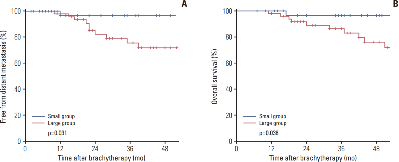
Distant metastases were diagnosed in 12 patients, and one patient died. The 3-year free from distant metastasis (FFDM) rate was 84%, and the OS was 90% for all patients. The FFDM and OS rates were also significantly higher in patients in the small group (3-year FFDM, 97% vs. 76%; p=0.031 and 3-year OS, 97% vs. 72%; p=0.036) (Fig. 3).
3. Eye preservation
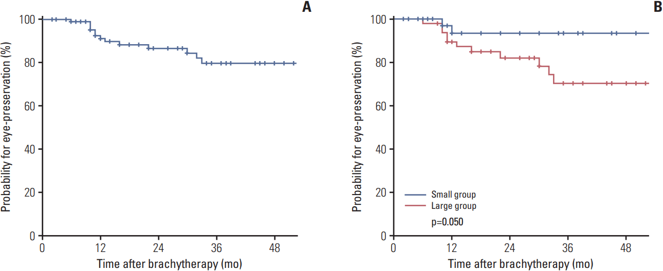
Among the 88 patients, 13 eventually underwent enucleation (12 patients for local recurrence and one patient for a complication), resulting in a 3-year actuarial eye-preservation rate of 80%. In the small group, only two patients underwent enucleation because of tumor progression, giving a 3-year eye-preservation rate of 94%. The eye-preservation rate was significantly higher in this group when compared with the large group (3-year eye-preservation rate, 94% vs. 70%, respectively; p=0.050) (Fig. 4).
4. Toxicities
The total rate of complications requiring further treatment was only 7% (n=6). The types, number of cases, and interval from brachytherapy are shown in Table 2. Two patients with glaucoma received medical treatment (mannitol, avastin), and one of them eventually received enucleation due to tumor progression, while the other underwent the Seton operation. Other complications, including vitreous hemorrhage and retinal detachment, were tolerable with proper intervention. One patient in the large group who had a tumor with a height of 9 mm and received TTT after brachytherapy wanted to undergo enucleation for phthisis 10 months after brachytherapy. The dose to the sclera of this patient exceeded 1,000 Gy.
Among the 63 patients with a pre-treatment visual acuity greater than 0.10, 39 (62%) showed a deterioration of visual acuity in the treated eye to < 0.10, which was considered to be legal blindness. The 3-year functional eye-preservation rate was 58%, and 50% of tumors were close to the macula or optic disc in these patients.
5. Prognostic factors
Cox regression analysis was used to evaluate prognostic factors for eye-preservation, distant metastasis, and OS (Table 3). The following potential prognostic variables were examined in the Cox proportional hazards models: age (< 50 years vs. ≥ 50 years), initial visual acuity, tumor height (< 6 mm vs. ≥ 6 mm), basal diameter, tumor location, and local treatment. The significant prognostic factor for eye-preservation was tumor height ≥ 6 mm (hazard ratio, 9.560; 95% confidence interval, 1.235 to 73.984, p=0.042), while it was not significantly associated with distant metastasis or OS (p=0.135 and p=0.157, respectively). Other characteristics including age, initial visual acuity, basal diameter, and tumor location did not affect the patients’ prognosis.
Discussion
In this study, we showed favorable local tumor control and eye-preservation rates, especially in patients in the small group (3-year local control, 94%; eye-preservation rate, 94%). These results are comparable to those reported by other groups for small or medium-sized tumors [12].
Although enucleation has been the most common primary treatment for large choroidal melanomas, previous studies showed that 125I brachytherapy could be a possible alternative treatment with regard to survival and local tumor control [13,14]. However, few studies have investigated the use of beta-radiation emitting 106Ru to treat large choroidal melanomas. 106Ru may not be effective because of its limited depth of penetration, which could prevent it from reaching the apex of thick tumors [15]. When compared with 125I, 106Ru has a threefold faster radiation dose fall-off rate. The effective amount of radiation of 80-100 Gy reaches less than 7 mm when a 106Ru eye plaque with 1,000 Gy to the outer surface of sclera is applied. Thus, a thicker tumor might receive an insufficient radiation dose to its apex. On the other hand, when the same radiation dose is delivered to the apex, the base receives up to three times more radiation with 106Ru than with 125I, which might cause extensive damage to the sclera. Based on these findings, most patients in the large group (96%) received additional local treatment with TTT, tumor excision or both.
Tumor necrosis caused by TTT is induced to a depth of 3 mm from the apex [16]. The effectiveness of combined use of 106Ru brachytherapy and TTT has been demonstrated in several studies. As with TTT, the apex of the tumor is treated up to 3 mm, while with brachytherapy, the highest dose is administered to the tumor base [10,17,18]. In addition, among patients in the large group (height ≥ 6 mm), local excision of the tumor was performed in 46% of patients. In such cases, the prescription point was determined to be 2 mm from the outer surface of the sclera. Augsburger et al. [19] previously reported treatment of choroidal melanomas with extrascleral extension by surgical excision of the extrascleral nodule followed immediately by plaque radiotherapy of the intraocular tumor. They demonstrated the efficacy of a combination of local excision and brachytherapy for the treatment of choroidal melanomas showing that brachytherapy alone was not sufficient. Adding these local treatments to 106Ru brachytherapy could increase local control in patients with tumor heights exceeding 6 mm, which is generally regarded as the maximum thickness suitable for 106Ru brachytherapy. Furthermore, it is expected that the radiation dose to the scleral surface will need to be reduced to decrease the risk of radiation-induced toxicities.
Choroidal melanomas are relatively resistant to radiation, and the brachytherapy dose needed to eradicate this tumor is associated with a considerable risk of radiation damage to ocular structures. In our analysis, the actuarial rate of radiation related side effects that require further treatment was 7%. One of these patients underwent enucleation for persistent phthisis after brachytherapy. This patient received a high dose of radiation to the sclera exceeding 1,000 Gy with a high dose rate (dose rate to sclera, 502.9 cGy/hr). Overall, 62% of patients who had effective visual function upon initial diagnosis eventually experienced complete loss of their visual acuity at their last follow-up, which is comparable to the results from other large series [20,21]. Half of these patients had a tumor close to the macula or the optic disc. Radiation induces occlusive vasculopathy with neovascularization and retinal edema, resulting in deterioration of visual acuity that continues for years after treatment. Increased doses and dose rates to the macula and the optic disc are associated with poorer visual outcomes [22].
According to our data, 80% of patients initially treated with brachytherapy could be spared from enucleation. As expected, the eye-preservation rate of the large group was lower than that of the small group (3-year eye-preservation rate, 70% vs. 94%; p=0.047). Additionally, the overall outcome of patients in our large group was lower than that of Caucasian patients, who showed a 70%-90% eye-preservation rate after 125I brachy-therapy for large melanomas and a 82%-88% rate after 106Ru brachytherapy for any sized tumors. Although 70% of patients in the large group were rescued from enucleation, their oncologic outcome was still not satisfactory (3-year FFDM, 76%; 3-year OS, 72%). This could be explained by the distinct nature of the tumor in Asian patients [23].
For these patients, other therapeutic approaches, including proton therapy, which can access the full thickness of a large tumor, may improve local control [24,25]. However, an effective method for reducing metastasis is still lacking, and survival rates for these patients do not differ significantly between treatment methods, including brachytherapy, enucleation, and GKS [1,13]. Hawkins reported that no effective treatment for metastatic ocular melanoma has been found during the process of organizing Collaborative Ocular Melanoma Study trials [26]. However, a recent study reported the safety and efficacy of immuno-therapy with anti–CTLA-4 antibody, an effective treatment for metastatic cutaneous melanoma, for treatment of metastatic uveal melanoma [27]. Based on the results of the present and previous studies, further investigation is warranted. Moreover, early detection of distant metastases at the time of diagnosis is essential to avoiding unnecessary local treatment and to evaluation of potential adjunctive treatments.
In interpreting our data, the limitations of retrospective analysis, including patients and treatment selection, must be considered. Although the absolute indication for brachytherapy with additional local treatment is unclear, use of this treatment seems to reliably be recommended for the improvement of local control. Another limitation lies in the difficulty of evaluating toxicities, including determining whether changes in the retina were attributed to radiation alone or to tumor growth as well, and if these changes may have existed at the time of initial diagnosis. Toxicity analysis included only the patients requiring further treatment in this study; therefore, the incidence of toxicities cannot be applied directly to all patients with choroidal melanomas treated with 106Ru brachytherapy. As the cohort is relatively small, there were limitations in conducting a complete statistical analysis. Additionally, it is necessary to verify the prognostic factors identified in our data. Nevertheless, the strengths of our study are that our patients received relatively homologous treatments based on their tumor size, and that they were performed in a single institution.